Physiology
Textbooks
Boundless Anatomy and Physiology
Physiology Textbooks Boundless Anatomy and Physiology
Physiology Textbooks
Physiology

Chapter 7

Skeletal System: Parts of the Skeleton

Book Version 29
By Boundless
Boundless Anatomy and Physiology
Physiology
by Boundless
View the full table of contents
Section 1
The Skull
Thumbnail
General Features and Functions of the Skull

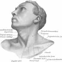
The human skull is the part of the skeleton that supports the structures of the face and forms a cavity for the brain.

Thumbnail
Cranial Bones

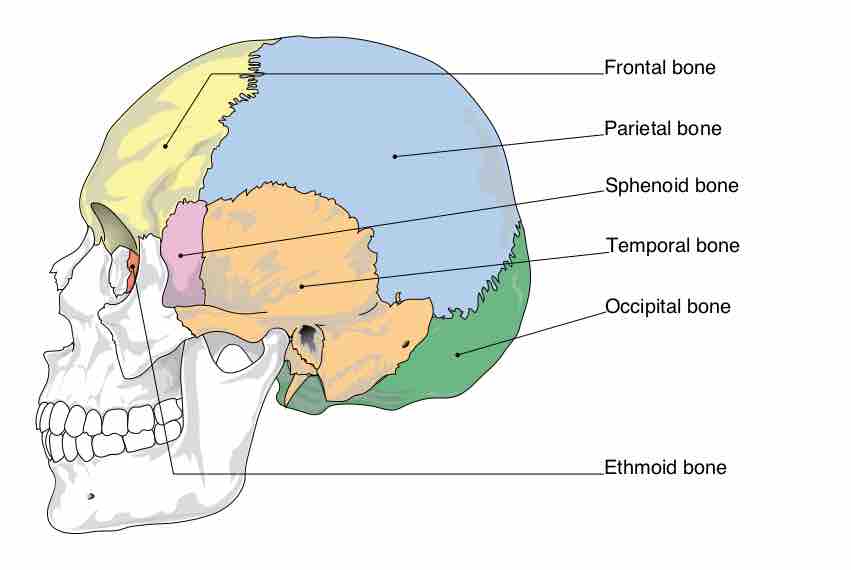
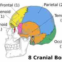
The neurocranium is comprised of eight bones: occipital, two temporal bones, two parietal bones, sphenoid, ethmoid, and the frontal bone.

Thumbnail
Facial Bones

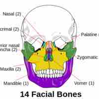
The viscerocranium (face) includes these bones: vomer, 2 inferior nasal conchae, 2 nasals, maxilla, mandible, palatine, 2 zygomatics, and 2 lacrimals.

Orbits

The orbit is the cavity or socket of the skull in which the eye and its appendages are situated.

Thumbnail
Foramina

The human skull has numerous holes known as foramina through which cranial nerves, arteries, veins, and other structures pass.

Thumbnail
Sutures

A suture is a type of fibrous joint (or synarthrosis) that only occurs in the skull (or cranium).

Thumbnail
Paranasal Sinuses

The paranasal sinuses (four, paired, air-filled spaces) surround the nasal cavity, and are located above and between the eyes, and behind the ethmoids.

Thumbnail
Fontanels

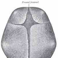
A fontanelle (or fontanel) is an anatomical feature on an infant's skull.

Section 2
The Hyoid Bone
Thumbnail
The Hyoid Bone

The hyoid, a horseshoe-shaped bone situated in the anterior of the neck, between chin and thyroid, aids tongue movement and swallowing.

Section 3
The Vertebral Column
Thumbnail
General Characteristics of the Spine

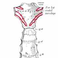
The spine is made of vertebrae that link together to protect the spinal cord.

Thumbnail
Parts of a Vertebra

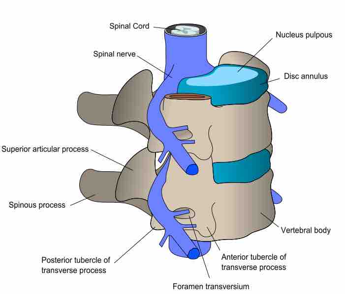
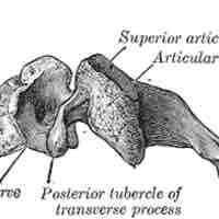
A vertebra consists of two parts: an anterior segment, or the vertebral body; and a posterior part, or the vertebral (neural) arch.

Thumbnail
Regional Vertebral Characteristics

The vertebrae of the spinal column are divided into five regions: cervical, thoracic, lumbar, sacral, and coccyx.

Section 4
The Thorax
Thumbnail
Thoracic Cage: Sternum

The sternum or breastbone is a long, flat, bony plate connected to the rib bones via cartilage that forms the anterior section of the rib cage.

Thumbnail
Thoracic Cage: Ribs

The ribs are long, curved bones that protect the lungs, heart, and other organs of the thoracic cavity.

Section 5
The Shoulder
Thumbnail
Clavicle

The clavicle or collar bone is a long, curved bone on the upper portion of the shoulder that connects with the scapula and the sternum.

Thumbnail
Scapula

The scapula, or shoulder bone, is a flat, triangular bone that connects to the humerus and the clavicle.

Section 6
The Upper Limb
Thumbnail
Humerus (The Upper Arm)

The bone forming the upper arm is the humerus.

Thumbnail
Ulna and Radius (The Forearm)

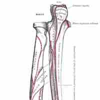
The forearm contains two bones, the radius and the ulna.

Thumbnail
Carpals, Metacarpals, and Phalanges (The Hand)

Each hand consists of 27 bones, divided between the wrist bones (carpals), the palm bones (metacarpals), and the finger bones (phalanges).

Section 7
The Hip
Thumbnail
Ilium

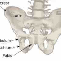
The ilium is the uppermost and largest bone of the pelvis.

Thumbnail
Ischium

The ischium forms the lower and posterior portion of the hip bones of the pelvis.

Thumbnail
Pubis

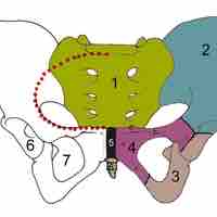
The pubis is the lowest and most anterior portion of the hip bones of the pelvis.

Thumbnail
False and True Pelves

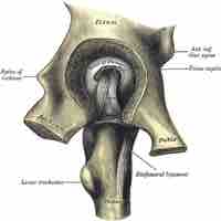
The false (greater) pelvis is larger and superior to the true (lesser) pelvis where the pelvic inlet is located.

Comparison of Female and Male Pelves

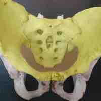
The female pelvis has evolved to its maximum width for childbirth and the male pelvis has been optimized for bipedal locomotion.

Thumbnail
Pelvic Structure and Childbearing

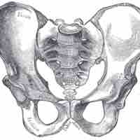
The human pelvis has evolved to be narrow enough for efficient upright locomotion, while still being wide enough to facilitate childbirth.

Section 8
The Lower Limb
Thumbnail
Femur (The Thigh)

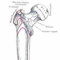
The femur—the bone of the upper leg—is the longest bone in the human body and one of the strongest.

Thumbnail
Patella (The Knee)

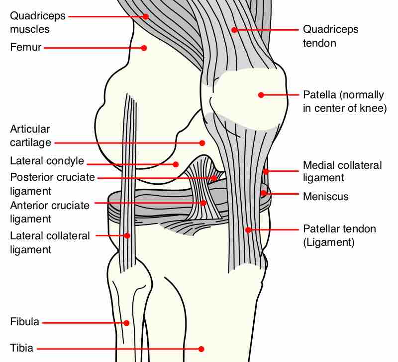
The patella (knee cap) is the bone between the fibula and femur.

Thumbnail
Tibia and Fibula (The Leg)

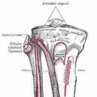
The tibia and the smaller fibula bones comprise the lower leg and articulate at the knee and ankle.

Thumbnail
Tarsals, Metatarsals, and Phalanges (The Foot)

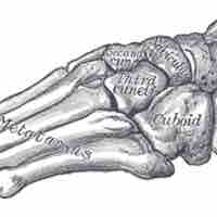
The human ankle and foot bones include tarsals (ankle), metatarsals (middle bones), and phalanges (toes).

Thumbnail
Arches of the Feet

The arches of the foot are formed by the tarsal and metatarsal bones; they dissipate impact forces and store energy for the subsequent step.

You are in this book
Boundless Anatomy and Physiology by Boundless
Previous Chapter
Chapter 6
Skeletal System
  • Overview of the Skeletal System
  • Cartilage
  • Introduction to Bone
  • Bone Formation
  • Bone and Calcium
Current Chapter
Chapter 7
Skeletal System: Parts of the Skeleton
  • The Skull
  • The Hyoid Bone
  • The Vertebral Column
  • The Thorax
  • The Shoulder
and 3 more sections...
Next Chapter
Chapter 8
Joints
  • Classification of Joints
  • Fibrous Joints
  • Cartilaginous Joints
  • Synovial Joints
Subjects
  • Accounting
  • Algebra
  • Art History
  • Biology
  • Business
  • Calculus
  • Chemistry
  • Communications
  • Economics
  • Finance
  • Management
  • Marketing
  • Microbiology
  • Physics
  • Physiology
  • Political Science
  • Psychology
  • Sociology
  • Statistics
  • U.S. History
  • World History
  • Writing

Except where noted, content and user contributions on this site are licensed under CC BY-SA 4.0 with attribution required.