Physiology
Textbooks
Boundless Anatomy and Physiology
Skeletal System: Parts of the Skeleton
The Vertebral Column
Physiology Textbooks Boundless Anatomy and Physiology Skeletal System: Parts of the Skeleton The Vertebral Column
Physiology Textbooks Boundless Anatomy and Physiology Skeletal System: Parts of the Skeleton
Physiology Textbooks Boundless Anatomy and Physiology
Physiology Textbooks
Physiology
Concept Version 11
Created by Boundless

Regional Vertebral Characteristics

The vertebrae of the spinal column are divided into five regions: cervical, thoracic, lumbar, sacral, and coccyx.

Learning Objective

  • Distinguish the differing types of vertebrae


Key Points

    • The upper three regions of the vertebral column are articulating vertebrae grouped under the names cervical, thoracic and lumbar, according to the regions they occupy.
    • Vertebrae are given an alphanumeric descriptor, with the initial letter derived from the region they are located in followed by a digit that increases moving down the region. For example, the most superior cervical vertebrae is termed C1 and the most inferior C7, which is then followed by the T1 vertebrae of the thoracic region.
    • The two lower most regions of vertebrae are the sacrum and the coccyx.

Full Text

The vertebrae comprising the spinal column can be divided into five regions, based on the five varying curvatures of the spine. The upper three regions of the spinal column are termed the cervical, thoracic, and lumbar; they contain individually jointed vertebrae. The two lower regions—the sacrum and coccyx, or tailbone—are formed from fused vertebrae.

Upper Vertebrae

Vertebrae are given an alphanumeric descriptor, with the initial letter derived from the region they are located in followed by a digit that increases moving down the region. For example, the most superior cervical vertebrae is termed C1 and the most inferior C7, which is then followed by the T1 vertebrae of the thoracic region.

The cervical region of the spine is the most superior and contains seven small vertebrae. The main function of the cervical region is to facilitate attachment of the skull to the spine, protect the spinal cord over the exposed neck and shoulder region, and support the body.

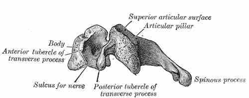
Cervical vertebra, lateral view

The lateral view of a typical cervical vertebra. 

This is a drawing of a lateral view of a typical cervical vertebra. It calls out these features of the vertebral body: the superior articular surface, the articular pillar, spinous process, posterior tubercle of transverse process, sulcus for nerve, and the anterior tubercle of transverse process.

The twelve thoracic vertebrae are located inferiorly to the cervical region. They are larger than the cervical vertebrae and increase in size moving inferiorly to the lumbar region.
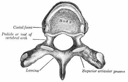
Thoracic vertebra

Image of a typical thoracic vertebra. 

This is a black and white drawing that calls out these features of a typical thoracic vertebra: the costal fovea, pedicle or root of the vertebral arch, lamina, and superior articular process.

The five lumbar vertebrae are the largest vertebral bones and increase in size when moving inferiorly. The lumbar vertebrae play a key role in supporting the body and facilitating locomotion.
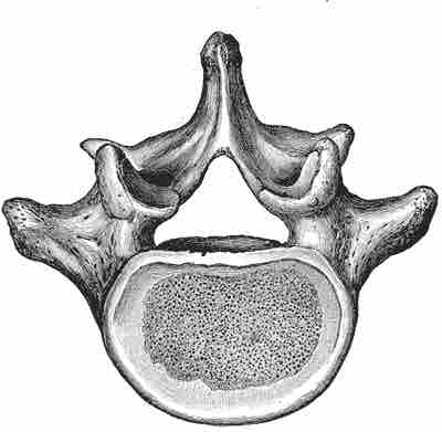
Lumbar vertebra

Image of typical lumbar vertebra.

This is a drawing showing a cross section of a typical lumbar vertebra.

Lower Vertebrae

During childhood the five vertebrae of the sacral region are distinct. In adulthood the five bones fuse to form the sacrum, although it is still often divided into regions termed S1–S5 based on the formation of the original individual bones. The sacrum functions to support the body and protect organs of the pelvis and lower back.

The final region of the spine is the coccyx, or tailbone. As with the sacrum, the coccyx is formed from several vertebrae that have fused together. 

As it’s alternative name suggests, the coccyx forms the basis of a tail that has been lost in humans, although it is incorrect to think of it as a vestigial structure since is a key attachment point for many muscles and ligaments and plays a key role in supporting the body while sitting.
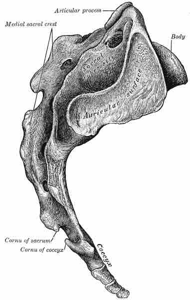
Coccyx

Lateral view of coccyx shown beneath the sacrum.

This is a drawing of a coccyx, shown from beneath the sacrum so its tail-like form is displayed.
[ edit ]
Edit this content
Prev Concept
Parts of a Vertebra
Thoracic Cage: Sternum
Next Concept
Subjects
  • Accounting
  • Algebra
  • Art History
  • Biology
  • Business
  • Calculus
  • Chemistry
  • Communications
  • Economics
  • Finance
  • Management
  • Marketing
  • Microbiology
  • Physics
  • Physiology
  • Political Science
  • Psychology
  • Sociology
  • Statistics
  • U.S. History
  • World History
  • Writing

Except where noted, content and user contributions on this site are licensed under CC BY-SA 4.0 with attribution required.