Physiology
Textbooks
Boundless Anatomy and Physiology
Skeletal System: Parts of the Skeleton
The Vertebral Column
Physiology Textbooks Boundless Anatomy and Physiology Skeletal System: Parts of the Skeleton The Vertebral Column
Physiology Textbooks Boundless Anatomy and Physiology Skeletal System: Parts of the Skeleton
Physiology Textbooks Boundless Anatomy and Physiology
Physiology Textbooks
Physiology
Concept Version 13
Created by Boundless

General Characteristics of the Spine

The spine is made of vertebrae that link together to protect the spinal cord.

Learning Objective

  • Describe the purpose of the vertebral column and its development from fetus to toddler


Key Points

    • The main functions of the vertebral bones are for structure (posture) and protection of the spinal cord.
    • The spine is split into five regions: the cervical, thoracic, lumbar, sacral, and coccyx.
    • The vertebrae of the sacrum and coccyx are fused, but those of the cervical, thoracic, and lumber regions are free to articulate.
    • Viewed laterally, the vertebral column presents several curves that correspond to the column's different regions—the cervical, thoracic, lumbar, and sacral.

Terms

  • vertebrae

    The bones that make up the spinal column.

  • vertebral column

    The series of vertebrae that protect the spinal cord; the spinal column.


Full Text

The vertebral column (also known as the backbone or spine), is a tall, thin organ located dorsally that extends from the base of the spine to the pelvis. It protects the spinal cord and provides a key attachment point for numerous muscle groups. 

There are 33 vertebrae in the human spine that are split into four regions that correspond to the curvature of the spine; the cervical, thoracic, lumbar, sacrum, and coccyx. The vertebrae of the sacrum and coccyx are fused, but those of the cervical, thoracic and lumbar regions are separated by intervertebral discs. 

Vertebrae are given an alphanumeric descriptor, with the initial letter derived from the region they are located in followed by a digit; the digit increases moving down the region. For example, the most superior cervical vertebra is termed C1 and the most inferior C7, which is then followed by the T1 vertebrae of the thoracic region.

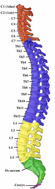
Human vertebral column

The vertebral column has 33 bones. Each color represents a section of the column.

This is a profile view of the spine with its four regions identified. These are called, from the top of the spine to its base, the cervical, thoracic, lumbar, and pelvic regions.

Viewed laterally the vertebral column presents several curves that correspond to the different regions of the column. These are called the cervical, thoracic, lumbar, and pelvic regions.

  • The cervical curve covers the region between vertebrae C1 and T2, it is the least marked of all the spinal curves.
  • The thoracic curve covers the region between vertebrae T2 and T12.
  • The lumbar curve covers the region between vertebrae T12 and L5 and is more marked in the females than in males due to differences in pelvic structure.
  • The sacral curve begins at the sacrovertebral articulation, and ends at the point of the coccyx.

The thoracic and sacral curves are termed primary curves because they alone are present during fetal life. The cervical and lumbar curves are secondary curves that are developed after birth; the former when the child is able to maintain an upright posture, the latter when the child begins to walk.

[ edit ]
Edit this content
Prev Concept
The Hyoid Bone
Parts of a Vertebra
Next Concept
Subjects
  • Accounting
  • Algebra
  • Art History
  • Biology
  • Business
  • Calculus
  • Chemistry
  • Communications
  • Economics
  • Finance
  • Management
  • Marketing
  • Microbiology
  • Physics
  • Physiology
  • Political Science
  • Psychology
  • Sociology
  • Statistics
  • U.S. History
  • World History
  • Writing

Except where noted, content and user contributions on this site are licensed under CC BY-SA 4.0 with attribution required.