The tibia and fibula are the two bones of the lower leg. The tibia is located medially to the fibula and is much larger. Both are bound together with the interosseous membrane.
The leg
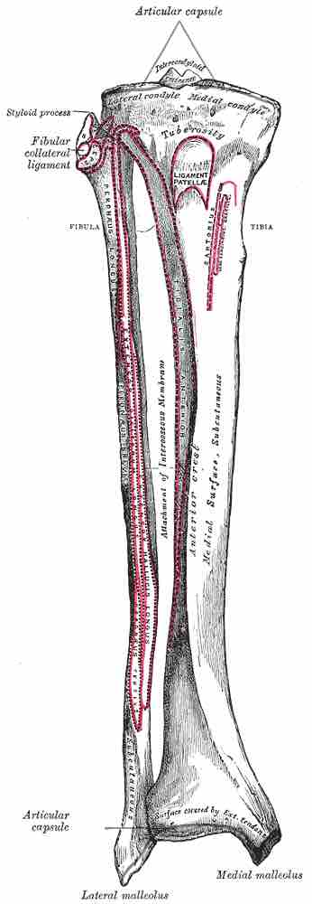
Tibia and fibula in anatomical position with parts labeled.
The Tibia
The tibia, or shin bone, spans the lower leg, articulating proximally with the femur and patella at the knee joint, and distally with the tarsal bones, to form the ankle joint. It is the major weight-bearing bone of the lower leg.
Proximally, there are five key features of the tibia:
- It widens and forms two condyles—the lateral and medial—that articulate with the condyles of the femur.
- Between the two condyles is the intercondylar fossa, a small grove, into which two intercondylar tubercles sit. Numerous internal ligaments of the knee joint attach to these tubercles and strengthen it significantly.
- On the anterior surface of the proximal region and inferiorly to the condyles is the tibial tuberosity to which the patella ligament attaches.
- The shaft of the tibia is triangular and the soleus muscle, which gives the calf its characteristic shape, originates on the posterior surface.
- Distally, the tibia also widens to aid with weight bearing and it displays two key features. The medial malleolus is a bony projection that articulates with the tarsal bones to form the ankle joint. Laterally, there is the fibular notch that articulates with the fibula.
The Fibula
The fibula also spans the lower leg, although proximally it does not articulate with the femur or patella. It serves more as an attachment point for muscles rather than a weight-bearing bone.
Proximally, the fibula head articulates with the lateral condyle of the tibia, and the biceps femoris attaches to the fibula head. As with the tibia, the shaft of the fibula is triangular and numerus muscles are involved in the extension and flexion of the foot. These muscles originate from the fibula's surface and include the extensor digitorum longus, soleus, and flexor hallucis longus, among others.
Distally, the fibula forms the lateral malleolus, which is more prominent than the medial malleolus of the tibia. It also articulates with the tarsal bones to form the ankle joint.