wikiHow is a “wiki,” similar to Wikipedia, which means that many of our articles are co-written by multiple authors. To create this article, 11 people, some anonymous, worked to edit and improve it over time.
This article has been viewed 59,540 times.
Learn more...
This guide will provide a description and example of how to draw the human respiratory system. First by outlining the lungs, bronchi, and trachea, then detailing additional components and accessory structures. By the end of this wikiHow, you will be proficient in drawing a medically accurate human respiratory system and be ready to ace your exams!
Steps
Drawing Basic Respiratory Structures
-
1Select a dynamic color scheme. For this activity, you will need 1-2 low contrast colors and 9 highlighting colors. There are several medical illustration standards to keep in mind, such as using blue for oxygenated blood flow and red for deoxygenated blood flow. Beyond those standards, it’s up to you how detailed you need your illustration to be.
-
2Select a reference photo. Choose a reference photo online or from your textbook to follow along with.Advertisement
-
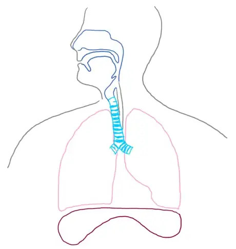
3Draw the frame for the diagram. Outline the thoracic cavity, neck, and head. You will want this to be large enough for you to draw more detailed structures inside. For a small diagram, outline the thoracic cavity to be no smaller than 3 in (8 cm) wide to ensure clarity. Do this with a low contrast color, so you can use more vibrant colors to highlight structures.
-
4Illustrate the lungs. Draw two rounded, somewhat triangular shapes in the thoracic cavity with the base on the nipple-line and the apex at the clavicle. These are the lungs! These triangles should be angled upward at the base in the medial direction. This distinction is important because the diaphragm is located below the lungs and occupies this space.
- Additionally, a notch should be placed in the left lung which accommodates for the heart (part of the cardiovascular system, not included in this illustration).
-
5Outline the trachea and bronchi trunk. Using your lungs as a reference, draw an upside-down Y shape centered at the top quarter of the lungs and leading upward to the neck. The center point should be at the suprasternal notch, and the long part of the Y should extend to the Adam’s apple. This Y shape is the two bronchus trunks and the trachea. These structures are cylindrical with notches similar to what you would see on a tin of soup; adding this detailing is beneficial to your viewer because it allows them to better understand the physiology of the structure.
-
6Sketch the air passageways. Continue the sketch of the trachea to the rest of the neck up to the mouth. Now curve this structure through the mouth and to the lips. The section you’ve just drawn is the larynx leading into the oropharynx. The nasal passageway, or nasopharynx, begins at the nostrils and connects to the oropharynx at the mandibular angle.
Adding Accessory Structures and Details
-
1Include landmark structures. Parallel to the larynx and oropharynx is the esophagus (part of the digestive system) but should be included in your illustration as a low-contrast color. At the mandibular angle, a small “flap” should be noted; this is the epiglottis, which covers the trachea when swallowing to prevent debris from entering the lungs.
-
2Sketch the diaphragm. Draw a flat kidney bean shape following the outline of the base of the lungs. The diaphragm is a muscle that contracts during inhalation and relaxes during exhalation to modify pressure in the lungs.
-
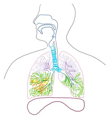
3Detail the lobes of the lungs. Segment the right lung into 3 curved sections (draw the oblique and horizontal fissures) and the left lung into 2 curved sections (draw the oblique fissure) as distributed below; these segments are called lobes.
-
4Outline the branching. Extend the branching of the bronchus in each lung into a secondary bronchus in each lobe. Then extend this secondary branching with smaller bronchioles. It is helpful to do each lobe branch in a new color to distinguish the branching pattern.
-
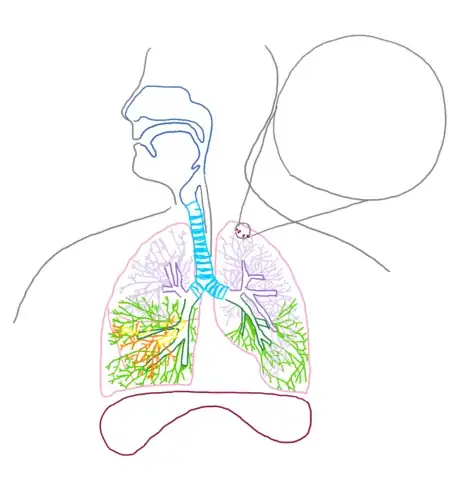
5Draw a microscope bubble. Attached to these bronchioles are alveoli, which appear as small grapes. Detail several of these and then extend a microscope box away from your diagram to illustrate this structure more clearly.
-
6Detail the alveoli. Redraw a segment of the bronchioles and attached alveoli in the microscope box. The bulbous “grape” like structures of the alveoli are called alveolar sacs, and the segment of branching immediately before the alveoli are called alveolar ducts.
- In addition to these structures, draw an overlay of the pulmonary artery (red) and pulmonary vein (blue) leading into the arteriole and venule capillary system.
-
7Label your completed diagram. Draw lines away from each structure to an open space using a ruler or straight edge. Clearly label each structure or region correctly. For more complex drawings, it is sometimes beneficial to label structures numerically and then provide an organized key.
Community Q&A
-
QuestionWhat is the work of the kidney?
 GB742Top AnswererThe kidneys are responsible for maintaining homeostasis in the body by filtering the blood and excreting excess fluids and electrolytes.
 -
QuestionWhat is the function of the epiglottis?
 GB742Top AnswererThe epiglottis is a flap which covers the trachea when swallowing to prevent food or liquid entering the lungs.
 -
QuestionWhat is number 14 in the bronchioles?
 GB742Top AnswererIn the case of this diagram, 14 indicates the alveolar sacs - this is the part of the lung where gas exchange occurs.
About This Article
To draw the human respiratory system, you'll need to outline the organs that make up the different parts and then add details to show how they function. Start by drawing an outline of the head, neck, and shoulders. Draw 2 rounded, somewhat triangular shapes in the cavity to represent the lungs and then add an upside-down Y shape for the trachea and the bronchi connected to the lungs. Then, sketch out the mouth and sinus passageways, and add the diaphragm beneath the lungs. From there, add lobes and branching to give the lungs more detail. You could also draw a microscope bubble and draw alveoli inside of it to illustrate the smaller structures inside of the lungs. Once you’ve finished your diagram, label all of the parts of the system. For tips about how to add an organized key to your diagram, read on!


