The gonad is the organ that makes gametes. The gonads in males are the testes and the gonads in females are the ovaries. Both gonads in males and females are endocrine glands.
The Ovaries
The ovary is a paired, ovum-producing, reproductive organ located in the lateral wall of the pelvis in a region called the ovarian fossa. The fossa usually lies beneath the external iliac artery and in front of the ureter and the internal iliac artery.
The ovaries are not attached to the fallopian tubes but to the outer layer of the uterus via the ovarian ligaments. Usually each ovary takes turns releasing eggs every month; however, if there was a case where one ovary was absent or dysfunctional then the other ovary would continue providing eggs to be released.
Ovaries secrete both estrogen and progesterone. Estrogen is responsible for the development of the secondary sex characteristics of human females at puberty and for the maturation and maintenance of the reproductive organs in their mature functional state. Progesterone functions with estrogen by promoting menstrual cycle changes in the endometrium. The ovaries also secrete testosterone, although at a much lower level than in males.
Progesterone and estrogen are secreted by granulosal cells, whereas testosterone is produced by thecal cells. Prior to ovulation, follicle-stimulating hormone is secreted by the granulosal cells that convert testosterone into estradiol.

Ovary
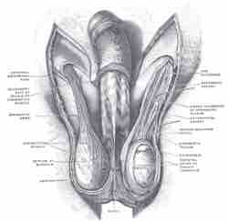
Blood supply of the human female reproductive organs. The left ovary is visible above the label ovarian arteries.
The Testes
The testes are the male reproductive gonads in humans. Like the ovaries, to which they are homologous, testes are components of both the reproductive system and the endocrine system. The primary functions of the testes are to produce sperm (spermatogenesis) and androgens, primarily testosterone.
The functions of the testicles are influenced by gonadotropic hormones, that are produced by the anterior pituitary. Luteinizing hormone results in testosterone release. The presence of both testosterone and follicle-stimulating hormone is needed to support spermatogenesis. Testosterone is secreted by Leydig cells, which are located between the seminiferous tubules.
The testes are located in the scrotum (a sac of skin between the upper thighs). In the male fetus, the testes develop near the kidneys, then descend into the scrotum just before birth. Each testis is about 1 1/2 inches long by 1 inch wide.
Testicles
Diagram of male (human) testicles.