The eye develops from the neural tube, the epidermis, and the periocular mesenchyme, which receives contributions from both the neural crest and mesoderm lineages. The organogenesis of the eye is an example of a developmental cascade of inductions with three different tissues contributing to its differentiations.
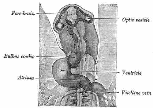
Neural tube: First, there is an outpocketing of the neural tube called optic vesicles . Development of the optic vesicles starts in the three week embryo from a progressively deepening groove in the neural plate called the optic sulcus. As this expands, the rostral neuropore (the exit of the brain cavity out of the embryo) closes and the optic sulcus and the neural plate becomes the optic vesicle.
Chick embryo head with optic vesicle
The eyes make their appearance before the closure of the anterior end of the neural tube. After the closure of the tube they are known as the optic vesicles.
Epidermis: The optic vesicles come into contact with the epithelum and induce the epidermis. The epithelium thickens to form the lens placode. The lens differentiates and invaginates until it pinches off from the epithelium. The lens acts as an inducer back to the optic vesicle to transform it into the optic cup and back to the epidermis to transform it into the cornea. The optic cup then delaminates into two layers: the neural retina and the retinal pigment epithelium.
Periocular mesenchyme: The periocular mesenchyme migrates inward during the formation of the optic cup. It is critical for the induction of the retinal pigment epithelium and the optic nerve. The mesenchyme contributes to the cornea, iris, ciliary body, sclera, and blood vessels of the eye.
Chordamesoderm induces the anterior portion of the neural tube to form the precursors of the synapomorphic tripartite brain of vertebrates, and it will form a bulge called the diencephalon. Further induction by the chordamesoderm will form a protrusion: the optic visicle. This visicle will be subsequently invaginated by means of further inductions from the chordamesoderm. The optic visicle will then induce the ectoderm that thickens (lens placode) and further invaginates to a point that detaches from the ectoderm and forms a neurogenic placode by itself. The lens placode is affected by the chordamesoderm making it invaginate and form the optic cup composed by an outer layer of neural retina and inner layer of pigmented retina that will unite and form the optic stalk . The pigmented retina is formed by rods and cones and composed of small cilia typical of the ependymal epithelium of the neural tube. Some cells in the lens vesicle will be fated to form the cornea and the lens vesicle will develop completely to form the definitive lens. Iris is formed from the optic cup cells.
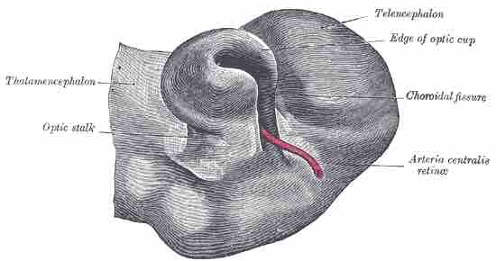
The Optic Stalk and Optic Cup
During embryonic development of the eye, the outer wall of the bulb of the optic vesicles becomes thickened and invaginated, and the bulb is thus converted into a cup, the optic cup.