Microbiology
Textbooks
Boundless Microbiology
Microbiology Textbooks Boundless Microbiology
Microbiology Textbooks
Microbiology

Chapter 3

Microscopy

Book Version 6
By Boundless
Boundless Microbiology
Microbiology
by Boundless
View the full table of contents
Section 1
Looking at Microbes
Thumbnail
Microbe Size

Microbes are often very small, even in comparison to microscopic cells from animals.

Thumbnail
Units of Measurement for Microbes

Measuring microbes presents challenges because they are very small, requiring indirect measures of microbes to understand them better.
Thumbnail
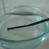
Refraction and Magnification

The underlying principal of a microscope is that lenses refract light which allows for magnification.
Thumbnail
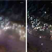
Magnification and Resolution

Magnification is the enlargement of an image; resolution is the ability to tell two objects apart.
Section 2
Light Microscopy
Thumbnail
Microscopy

Microscopes allow for magnification and visualization of cells and cellular components that cannot be seen with the naked eye.

Thumbnail
General Staining Methods

To properly visualize a microbe under a microscope, microbiologists use an array of stains to highlight cells and structures.

Section 3
Other Types of Microscopy
Thumbnail
Dark-Field Microscopy

Dark-field microscopes show a light silhouette of an organism against a dark background.
Thumbnail
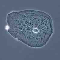
Phase-Contrast Microscopy

Phase-contrast microscopy visualizes differences in the refractive indexes of different parts of a specimen relative to unaltered light.
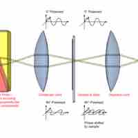
Thumbnail
Interference Microscopy

Interference microscopy is a variation of phase-contrast microscopy that uses a prism to split a light beam in two.
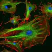
Thumbnail
Fluorescence Microscopy

Fluorescence microscopy is used to study specimens that are chemically manipulated to emit light.
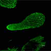
Thumbnail
Confocal Microscopy

The key to the confocal approach is the use of spatial filtering techniques to eliminate out-of-focus light from biological samples.
Thumbnail
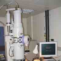
Electron Microscopy

Electron microscopy uses magnetic coils to direct a beam of electrons from a tungsten filament through a specimen and onto a monitor.
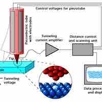
Thumbnail
Scanned-Probe Microscopy

Scanned-probe microscopy uses a fine probe rather than a light-beam or electrons to scan the surface of a specimen and produce a 3D image.
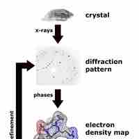
Thumbnail
X-Ray Diffraction Analysis

X-ray diffraction is a method that characterizes the structural composition of matter and using mathematical models.
You are in this book
Boundless Microbiology by Boundless
Previous Chapter
Chapter 2
Chemistry
  • Atomic Structure
  • Chemical Bonds
  • Chemical Reactions
  • Inorganic Compounds
  • Organic Compounds
and 2 more sections...
Current Chapter
Chapter 3
Microscopy
  • Looking at Microbes
  • Light Microscopy
  • Other Types of Microscopy
Next Chapter
Chapter 4
Cell Structure of Bacteria, Archaea, and Eukaryotes
  • Overview of Prokaryotic and Eukaryotic Cells
  • The Cytoplasmic Membrane of Prokaryotic and Eukaryotic Cells
  • Transport Across the Cell Membrane
  • Cell Walls of Prokaryotes
  • Specialized External Structures of Prokaryotes
and 5 more sections...
Subjects
  • Accounting
  • Algebra
  • Art History
  • Biology
  • Business
  • Calculus
  • Chemistry
  • Communications
  • Economics
  • Finance
  • Management
  • Marketing
  • Microbiology
  • Physics
  • Physiology
  • Political Science
  • Psychology
  • Sociology
  • Statistics
  • U.S. History
  • World History
  • Writing

Except where noted, content and user contributions on this site are licensed under CC BY-SA 4.0 with attribution required.