The ureters are tubes made of smooth muscle fibers that propel urine from the kidneys to the urinary bladder. In the adult, the ureters are usually 25–30 cm (10–12 in) long and 3–4 mm in diameter. The ureter is one of the essential organs of urinary tract that controls urine transport.
Ureter Structure and Function
The ureters are two tubes that are made out of smooth muscle and transitional epithelial tissues, which are a type of epithelial tissue that may either be columnar or squamous. Each kidney has its own ureter through which urine drains into.
The ureters are long tubes that have a few points of constriction, where obstructions are more common. The ureters receive a blood supply from a few different major arteries including the renal and illiac artery derivatives, and have a relatively dense nerve supply as well.
Muscles in the walls of the ureters send the urine in small spurts into the bladder, in a process called peristalsis. After the urine enters the bladder from the ureters, small folds in the bladder mucosa act like valves to prevent the backward flow of the urine; these are called the ureteral valves. The ureteral valves function similarly to the semilunar valves in the veins of the body, but are structurally different, consisting of transverse mucosal epithelial folds.
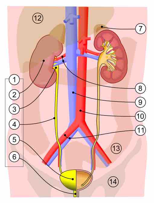
Human urinary system
1) Human urinary system. 2) Kidney. 3) Renal pelvis. 4) Ureter. 5) Urinary bladder. 6) Urethra (left side with frontal section). 7) Adrenal gland vessels. 8) Renal artery and vein. 9) Inferior vena cava. 10) Abdominal aorta. 11) Common iliac artery and vein with transparency. 12) Liver. 13) Large intestine. 14) Pelvis.
Ureter Pathology
Kidney stones and cancer are common diseases of the ureter. A kidney stone can move from the kidney and become lodged inside the ureter, which can block the flow of urine, as well as cause a sharp cramp in the back, side, or lower abdomen. The affected kidney could then develop hydronephrosis, should a part of the kidney become swollen due to blocked flow of urine.
Kidney stones are very common and are usually clumps of aggregated minerals that are most often found at the constriction points in the ureter. Ureter cancer is often due to a malignant transformation of of the transitional epithelial tissue, which is more vulnerable to developing cancer cells compared to other tissues.