Physiology
Textbooks
Boundless Anatomy and Physiology
Urinary System
Urine Transport, Storage, and Elimination
Physiology Textbooks Boundless Anatomy and Physiology Urinary System Urine Transport, Storage, and Elimination
Physiology Textbooks Boundless Anatomy and Physiology Urinary System
Physiology Textbooks Boundless Anatomy and Physiology
Physiology Textbooks
Physiology
Concept Version 14
Created by Boundless

Urinary Bladder

The urinary bladder is a hollow, muscular, and distendible or elastic organ that sits on the pelvic floor.

Learning Objective

  • Summarize the features and function of the urinary bladder


Key Points

    • The bladder is made of transitional epithelium and has a trigone.
    • A normal bladder empties completely upon a complete discharge, otherwise it is a sign that its elasticity is compromised.
    • When the bladder becomes half full, stretch receptors send nerve impulses to the spinal cord, which then sends a reflex nerve impulse back to the sphincter (muscular valve) at the neck of the bladder, causing it to relax and allow the flow of urine into the urethra.
    • The internal urethral sphincter is involuntary and the external sphincter is voluntary.
    • The bladder plays a minor role in temperature regulation.
    • The bladder has both mechanical and sensory innervation through the sympathetic and parasympathetic nervous systems.

Terms

  • urine

    A liquid excrement consisting of water, salts, and urea that is made in the kidneys then released through the urethra.

  • trigone

    A triangle-shaped part of an organ, such as the bottom of the bladder.

  • reflex

    An automatic response to a simple stimulus that does not require mental processing.


Full Text

The Urinary Bladder

The urinary bladder is a urine storage organ that is a part of the urinary tract. The bladder is a hollow, muscular, and elastic organ that sits on the pelvic floor. The bladder expands and fills with urine before it is discharged into the urethra during urination.

Bladder Anatomy

The bladder is a hollow, sac-like organ made of transitional epithelium, similar to the ureter that feeds into it. The ureters enter the bladder diagonally from its dorsolateral floor in an area called the trigone, which is a triangle-shaped anatomical region. The urethra exits at the lowest point of the triangle of the trigone.

There are two sphincters, or muscular valves, that separate the bladder from the urethra. The sphincters must open before the urine can flow into the urethra. The internal sphincter is under involuntary control and the external sphincter is under voluntary control.

Bladder Physiology

When the bladder fills with urine stretch receptors send nerve impulses to the spinal cord, which then sends a reflex nerve impulse back to the internal sphincter valve at the neck of the bladder that causes it to relax and allow the flow of urine into the urethra. The internal urethral sphincter is involuntary and controlled by the autonomic nerves. 

The bladder has a minor temperature regulation function since some heat may leave the body in the form of urine. A normal bladder empties completely upon a complete discharge, otherwise it is a sign that its elasticity is compromised; when it becomes completely void of fluid, it may cause a chilling sensation due to the rapid change of body temperature.

The urinary bladder usually holds 300–350 ml of urine. As urine accumulates, the walls of the bladder thin as it stretches, allowing the bladder to store larger amounts of urine without a significant rise in internal pressure of the bladder. 

The bladder receives motor innervation from both sympathetic fibers, most of which arise from the hypogastric plexuses and nerves, and parasympathetic fibers, which come from the pelvic splanchnic nerves and the inferior hypogastric plexus. Sensation from the bladder is transmitted to the central nervous system (CNS) via general visceral afferent fibers.
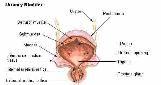
The urinary bladder

The urinary bladder is composed of several layers of tissue that facilitate urine storage and expulsion. The associated structures of the urinary and male reproductive tract are labelled. 

This is an illustration of the urinary bladder. It shows how the urinary bladder is composed of several layers of tissue that facilitate urine storage and expulsion. The associated structures of the urinary and male reproductive tract are labelled. These are the ureter, peritoneum, detrusor muscle, submucosa, mucosa, fibrous connective tissue, internal urethral orifice, external urethral orifice, rugae, ureteral opening, trigone, and the prostate gland.
[ edit ]
Edit this content
Prev Concept
Ureters
Urethra
Next Concept
Subjects
  • Accounting
  • Algebra
  • Art History
  • Biology
  • Business
  • Calculus
  • Chemistry
  • Communications
  • Economics
  • Finance
  • Management
  • Marketing
  • Microbiology
  • Physics
  • Physiology
  • Political Science
  • Psychology
  • Sociology
  • Statistics
  • U.S. History
  • World History
  • Writing

Except where noted, content and user contributions on this site are licensed under CC BY-SA 4.0 with attribution required.