Physiology
Textbooks
Boundless Anatomy and Physiology
Peripheral Nervous System
Motor Activity
Physiology Textbooks Boundless Anatomy and Physiology Peripheral Nervous System Motor Activity
Physiology Textbooks Boundless Anatomy and Physiology Peripheral Nervous System
Physiology Textbooks Boundless Anatomy and Physiology
Physiology Textbooks
Physiology
Concept Version 15
Created by Boundless

Peripheral Motor Endings

A neuromuscular junction exists between the axon terminal and the motor end plate of a muscle fiber where neurotransmitters are released.

Learning Objective

  • Describe the function of peripheral motor endings


Key Points

    • A neuromuscular junction is the junction between the axon terminal of a motor neuron and the plasma membrane of the motor end plate of a muscle fiber.
    • With the arrival of an action potential to the axon terminal, voltage-dependent calcium channels open, and calcium infuses into the cell. The influx of calcium ions causes the docking of acetylcholine-containing vesicles at the plasma membrane of the neuron and exocytosis into the synaptic cleft.
    • Acetylcholine is a neurotransmitter contained in the vesicles of the pre-synaptic neuron. It is released into the synaptic cleft and activates nicotinic acetylcholine receptors on the motor end plate, and causes local motor end plate depolarization, also known as the end plate potential (EPP).
    • The end plate potential propagates across the surface of the muscle fiber, causing the fiber to contract and continuing the process of excitation–contraction coupling.

Terms

  • axon

    A nerve fiber that is a long, slender projection of a nerve cell that conducts nerve impulses away from the body of the cell to a synapse.

  • presynaptic neuron

    The neuron that releases neurotransmitters into the synaptic cleft.

  • voltage-dependent calcium channels

    A group of voltage-gated ion channels found in excitable cells (e.g., muscle, glial cells, neurons, etc. ) with a permeability to the ion Ca2+.

  • excitation-contraction coupling

    This process is fundamental to muscle physiology, whereby the electrical stimulus is usually an action potential and the mechanical response is a contraction.

  • nicotinic acetylcholine receptor

    These are cholinergic receptors that form ligand-gated ion channels in the plasma membranes of certain neurons and on the postsynaptic side of the neuromuscular junction.

  • synaptic cleft

    A small space between neurons.


Full Text

A neuromuscular junction is the synapse or junction of the axon terminal of a motor neuron with the motor end plate, as shown in Figures 1 and 2. The highly excitable region of muscle fiber plasma membrane is responsible for initiation of action potentials across the muscle's surface, ultimately causing the muscle to contract. 

In vertebrates, the signal passes through the neuromuscular junction via the neurotransmitter acetylcholine.

Figure 1. Detailed view of a neuromuscular junction

Detailed view of a neuromuscular junction: 1) Presynaptic terminal; 2) Sarcolemma; 3) Synaptic vesicle; 4) Nicotinic acetylcholine receptor; 5) Mitochondrion.

This is a detailed view of a neuromuscular junction. The drawing identifies these parts in the junction: 1) Presynaptic terminal; 2) Sarcolemma; 3) Synaptic vesicle; 4) Nicotinic acetylcholine receptor; 5) Mitochondrion.

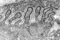
Figure 2. Neuromuscular junction

Electron micrograph showing a cross section through the neuromuscular junction. T is the axon terminal and M is the muscle fiber. The arrow shows junctional folds with basal lamina. Postsynaptic densities are visible on the tips between the folds. Scale is 0.3 µm.

This is an electron micrograph that shows a cross section through the neuromuscular junction. T labels the axon terminal and M labels the muscle fiber. The arrow shows junctional folds with basal lamina. Postsynaptic densities are visible on the tips between the folds. The scale is 0.3 µm.

Upon the arrival of an action potential at the presynaptic neuron terminal, voltage-dependent calcium channels open and Ca2+ ions flow from the extracellular fluid into the presynaptic neuron's cytosol. This influx of Ca2+ causes neurotransmitter-containing vesicles to dock and fuse to the presynaptic neuron's cell membrane, which results in the emptying of the vesicle's contents (acetylcholine) into the synaptic cleft; this process is known as exocytosis. 

Acetylcholine diffuses into the synaptic cleft and binds to the nicotinic acetylcholine receptors located on the motor end plate.

These receptors open to allow sodium ions to flow in and potassium ions to flow out of the muscle's cytosol, producing a local depolarization of the motor end plate, known as an end-plate potential (EPP). This depolarization spreads across the surface of the muscle fiber and continues the excitation–contraction coupling to contract the muscle. 

The action potential spreads through the muscle fiber's network of T-tubules, depolarizing the inner portion of the muscle fiber. The depolarization activates L-type, voltage-dependent calcium channels (dihydropyridine receptors) in the T-tubule membrane, which are in close proximity to calcium-release channels (ryanodine receptors) in the adjacent sarcoplasmic reticulum. 

As intracellular calcium levels rise, the motor proteins responsible for the contractile response are able to interact, as shown in Figure 3, to form cross-bridges and undergo shortening.

Clinical Example: Myasthenia gravis is an autoimmune disorder in which circulating antibodies block the nicotinic acetylcholine receptors on the motor end plate of the neuromuscular junction. This blockage of acetylcholine receptors causes muscle weakness, often first exhibiting drooping eyelids and expanding to include overall muscle weakness and fatigue.

The affects of myasthenia gravis illustrate the importance of effective and functioning neuromuscular junctions for communication between neurons and muscles to allow contraction and relaxation of muscle fibers.

Figure 3. Muscle contraction and actin–myosin interactions

Skeletal muscle contracts following activation by an action potential. The binding of acetylcholine at the motor end plate leads to intracellular calcium release and interactions between myofibrils to elicit contraction.

This diagram shows how muscular contraction is caused at the molecular level. A skeletal muscle contracts following activation by an action potential. The binding of acetylcholine at the motor end plate leads to intracellular calcium release and interactions between myofibrils to elicit contraction.
[ edit ]
Edit this content
Prev Concept
Sensory and Motor Tracts
Overview of Motor Integration
Next Concept
Subjects
  • Accounting
  • Algebra
  • Art History
  • Biology
  • Business
  • Calculus
  • Chemistry
  • Communications
  • Economics
  • Finance
  • Management
  • Marketing
  • Microbiology
  • Physics
  • Physiology
  • Political Science
  • Psychology
  • Sociology
  • Statistics
  • U.S. History
  • World History
  • Writing

Except where noted, content and user contributions on this site are licensed under CC BY-SA 4.0 with attribution required.