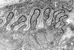
Figure 2. Neuromuscular junction
Electron micrograph showing a cross section through the neuromuscular junction. T is the axon terminal and M is the muscle fiber. The arrow shows junctional folds with basal lamina. Postsynaptic densities are visible on the tips between the folds. Scale is 0.3 µm.
This is an electron micrograph that shows a cross section through the neuromuscular junction. T labels the axon terminal and M labels the muscle fiber. The arrow shows junctional folds with basal lamina. Postsynaptic densities are visible on the tips between the folds. The scale is 0.3 µm.
Source
Boundless vets and curates high-quality, openly licensed content from around the Internet. This particular resource used the following sources: