Physiology
Textbooks
Boundless Anatomy and Physiology
Integumentary System
Cell Junctions
Physiology Textbooks Boundless Anatomy and Physiology Integumentary System Cell Junctions
Physiology Textbooks Boundless Anatomy and Physiology Integumentary System
Physiology Textbooks Boundless Anatomy and Physiology
Physiology Textbooks
Physiology
Concept Version 11
Created by Boundless

Tight Junctions

Tight junctions serve as selectively permeable seals in our body's internal and external surfaces.

Learning Objective

  • Describe the function of tight junctions


Key Points

    • Tight junctions are the closely associated areas of two cells whose membranes join together to form a virtually impermeable barrier to fluid.
    • Tight junctions perform vital functions—such as holding cells together—and form protective and functional barriers.
    • Tight junctions are composed of a branching network of sealing strands with each strand acting independently from the others.
    • The major types of proteins in junctions are the claudins and the occludins.
    • Each strand is formed from a row of transmembrane proteins embedded in both plasma membranes, with extracellular domains joining one another directly.

Terms

  • cell adhesion molecule

    Molecules that help cells stick to each other and to their surroundings. The proteins located on the cell surface bind with other cells or with the extracellular matrix (ECM).

  • cytoskeleton

    A cellular structure like a skeleton, contained within the cytoplasm.

  • blood-brain barrier

    A structure in the central nervous system (CNS) that keeps various substances found in the bloodstream out of the brain while allowing in the substances essential to metabolic function, e.g., oxygen.

  • Claudins

    Proteins that form the backbone of the tight junction strands.

  • epithelia

    The covering of internal and external body surfaces, where tight junctions are found.

  • zonula occludens

    Another name for tight junctions.


Full Text

Tight Junctions

Imagine a largely waterproof zipper connecting the sides of two different jackets.  That zipper is like a tight junction (TJ), also called an occluding junction. A TJ creates a small zone that occludes the extracellular space (the space between cells). 

This is why tight junctions are also called zonula occludens.  The word zonula comes from words that mean small zone or encircling belt, while occludens comes from the Latin word occludere, which means to close up.
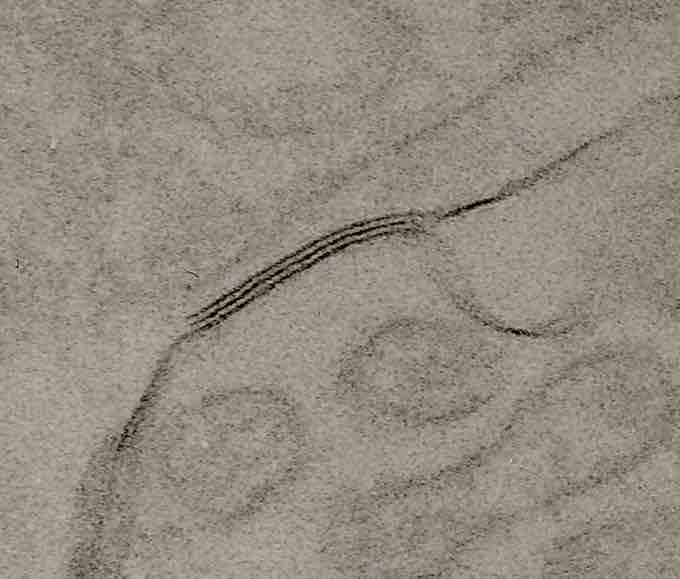
Tight Junction

An electron micrograph showing a tight junction in rat kidney tissue. The three dark lines of density correspond to the tight junction and the light lines in between correspond to the paracellular space.

This image is an electron micrograph showing a tight junction in rat kidney tissue. The three dark lines can be seen in the center of the image; these dark lines of density correspond to the tight junction and the light lines in between correspond to the paracellular space.

Location and Function

Tight junctions are virtually (but also partly selectively) impermeable seals that encircle cells and bind them together into leakproof sheets. In other words, the plasma membranes of adjacent cells essentially fuse together tightly in order to limit the leakage of various substances between the two cells.  

What can and cannot go through all depends on the substance’s size, charge, as well as the location and precise composition of the tight junctions in the part of the body in question.  

Tight junctions are located within our body’s epithelia.  Epithelia is the plural of epithelium. Epithelium is a word that refers to the covering of the body’s internal and external surfaces. This includes organs (such as skin), blood vessels, and cavities.

Thus, these tight junctions serve various functions, depending on what epithelium is in question. In the skin, they keep us somewhat watertight and help keep allergens out of our body. In the digestive system, they help prevent the leakage of digestive enzymes into our bloodstream. 

Tight junctions also serve as a structural support mechanism that help keep the epithelium together.
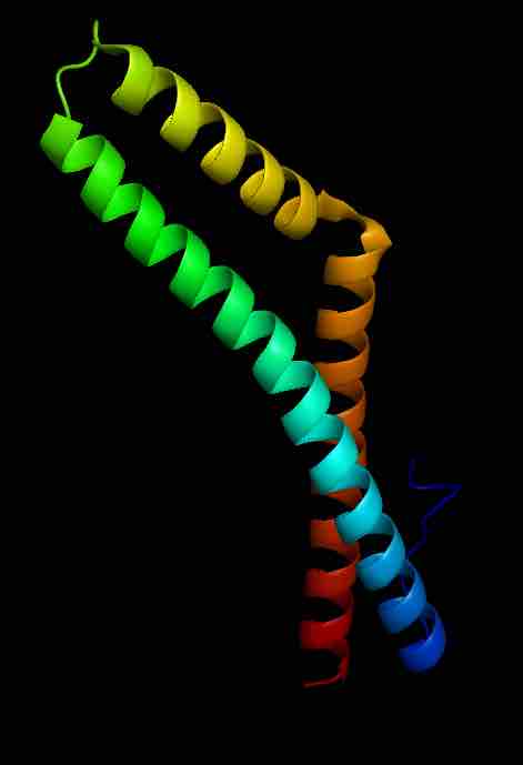
Occludin

Model of the protein structure of the coiled-coil domain of human occludin.

This is a computer-generated image of the protein structure of the coiled-coil domain of human occludin.

Composition

A tight junction—a kind of symmetrical cell junction—is composed of numerous important proteins that are either directly involved in its composition or intimately involved with connecting the tight junction to and between the cells in one way or another.  These proteins include:

  • Occludins, which maintain the barrier between adjacent cells.
  • Claudins, which form the backbone of tight junction strands.
  • Junctional adhesion molecules (JAMs) are immunoglobulin (antibody) proteins that help seal the intercellular space between two cells.
  • Zonula occludens (ZO) are proteins that help link the tight junction to each cell's internal skeleton (cytoskeleton).

The occludins and claudins are the major components of tight junction strands. When fully formed, a tight junction is not one, long, continuous seal.  Instead, it looks like a series of local seals joined together in a maze-like fashion.  
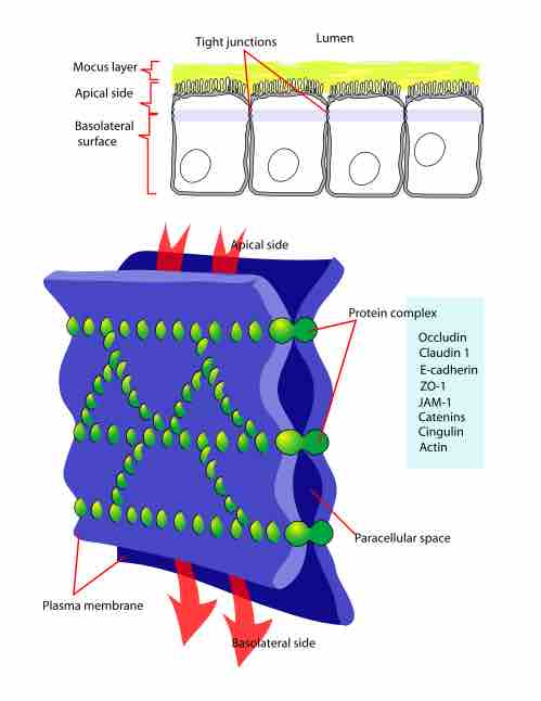
Tight junction

Diagram of tight junction components. 

This is a color diagram of tight junction components in a plasma membrane. It shows how tight junctions form seals that create paracellular spaces throughout the plasma membrane.
[ edit ]
Edit this content
Prev Concept
Skin Color
Adherens Junctions
Next Concept
Subjects
  • Accounting
  • Algebra
  • Art History
  • Biology
  • Business
  • Calculus
  • Chemistry
  • Communications
  • Economics
  • Finance
  • Management
  • Marketing
  • Microbiology
  • Physics
  • Physiology
  • Political Science
  • Psychology
  • Sociology
  • Statistics
  • U.S. History
  • World History
  • Writing

Except where noted, content and user contributions on this site are licensed under CC BY-SA 4.0 with attribution required.