Physiology
Textbooks
Boundless Anatomy and Physiology
Integumentary System
Cell Junctions
Physiology Textbooks Boundless Anatomy and Physiology Integumentary System Cell Junctions
Physiology Textbooks Boundless Anatomy and Physiology Integumentary System
Physiology Textbooks Boundless Anatomy and Physiology
Physiology Textbooks
Physiology
Concept Version 11
Created by Boundless

Adherens Junctions

Adherens junctions provide strong mechanical attachments between adjacent cells through the linkage of cytoplasmic face with cytoskeleton.

Learning Objective

  • Identify the characteristics of adherens junctions in epithelial tissues


Key Points

    • Adherens junctions are involved in a number of critical functions, including providing additional structural support. For example, they hold cardiac muscle cells tightly together as the heart expands and contracts.
    • Adherens junctions are built primarily from cadherins, whose extracellular segments bind to each other and whose intracellular segments bind to catenins. Catenins are connected to actin filaments.

Terms

  • cadherin

    Any of a class of transmembrane proteins important in maintaining tissue structure.

  • adherens junctions

    Protein complexes that occur at cell–cell junctions in epithelial tissues; they are usually more basal than tight junctions.

  • catenin

    Any of a class of proteins that have a role in cell adhesion.


Full Text

Adherens Junctions

Adherens junctions are also referred to as zonula adherens, intermediate junction, or as belt desmosomes.  Zonula means small zone or belt-like, and adherens refers to adhesion (sticking together).  As a result, the zonula adherens often runs like a belt around the entire cell in a continuous fashion, and it acts as an adhesion belt. 

Location and Function

This type of cell junction is located right below tight junctions and provides a strong bond between the sides of adjacent epithelial cell membranes.  While other junctions, like tight junctions, provide some support for and fusion of adjacent cells, their resistance to mechanical stress is relatively small compared to the much stronger adherens junctions.  

Structure and Composition

The zonula adherens is composed of several different proteins:

  • The actin microfilaments of the cytoskeleton (the internal skeleton of the cell). 
  • Anchor proteins, found inside each cell.  These are called alpha-catenin, beta-catenin, gamma-catenin (aka plakoglobin), vinculin, and alpha-actinin.  They link the actin microfilaments to the cadherins.
  • Cadherins, namely E-cadherin.  These are transmembrane adhesion proteins, whose main portions are located in the extracellular space.  

The extracellular part of one cell’s cadherin binds to the extracellular part of the adjacent cell’s cadherin in the space between the two cells.  Each cell’s cadherin molecule also contains a tail that inserts itself inside its respective cell.  

This intracellular (within the cell) tail then links up to catenin proteins to form the cadherin–catenin complex.  This complex binds to vinculin and alpha-actinin; these two proteins are what link the cadherin–catenin complex to the cell’s internal skeletal framework (the actin microfilaments). 

The extracellular portions of the cadherin molecules of adjacent cells are bonded together by calcium ions (or another protein in some cases).  This means that the functional as well as morphological integrity of the adherens junctions are calcium dependent. If you were to remove calcium from the equation, this type of cell junction would disintegrate as a result.  

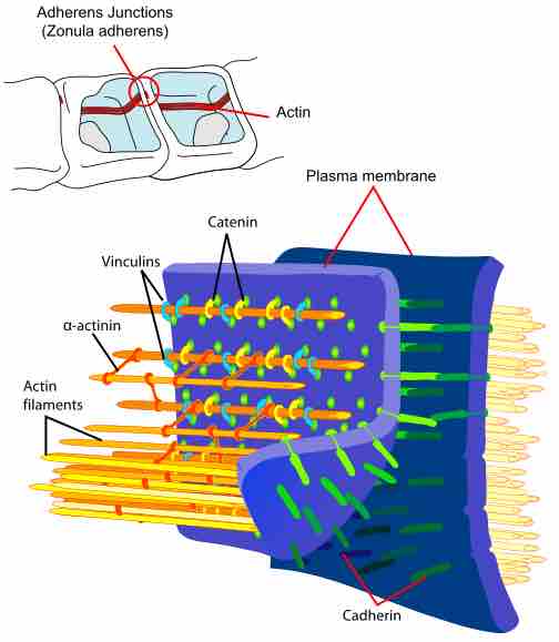
The structural proteins in an adherens junction 

These are the principal interactions of structural proteins at a cadherin-based plasma membrane adherens junction. Actin filaments are associated with adherens junctions in addition to several other actin-binding proteins. 

This illustration is two drawings of the principal interactions of structural proteins at a cadherin-based adherens junction. There are two pictures: one is a cut-away view of an adherens junctions with the actin filament identified. Actin filaments are associated with adherens junctions in addition to several other actin-binding proteins. The other is an exploded view of two plasma membranes that depict actin filaments as tubes that are joined to one another by bonds of a-actinin, and then joined to the plasma membrane by catenin and vinculins. The plasma membranes are joined together by cadherin protein.
[ edit ]
Edit this content
Prev Concept
Tight Junctions
Gap Junctions
Next Concept
Subjects
  • Accounting
  • Algebra
  • Art History
  • Biology
  • Business
  • Calculus
  • Chemistry
  • Communications
  • Economics
  • Finance
  • Management
  • Marketing
  • Microbiology
  • Physics
  • Physiology
  • Political Science
  • Psychology
  • Sociology
  • Statistics
  • U.S. History
  • World History
  • Writing

Except where noted, content and user contributions on this site are licensed under CC BY-SA 4.0 with attribution required.