Physiology
Textbooks
Boundless Anatomy and Physiology
Digestive System
The Stomach
Physiology Textbooks Boundless Anatomy and Physiology Digestive System The Stomach
Physiology Textbooks Boundless Anatomy and Physiology Digestive System
Physiology Textbooks Boundless Anatomy and Physiology
Physiology Textbooks
Physiology
Concept Version 11
Created by Boundless

Microscopic Anatomy of the Stomach

The layers of the stomach produce mucous to protect itself, enyzmes to break down the food for digestion, and muscles to 'churn' the food.

Learning Objective

  • Diagram the microscopic anatomy of the stomach


Key Points

    • The stomach walls are made of the following layers (inside to outside): mucosa, muscularis mucosa, submucosa, and muscularis externa.
    • The epithelium of the stomach forms deep pits (fundic or oxyntic glands) where chief cells produce pepsinogen, an inactive precursor of pepsin, which degrades proteins. The secretion of pepsinogen prevents self-digestion of the stomach cells.
    • Gastric acid kills most of the bacteria in food, stimulates hunger, and activates pepsinogen into pepsin; and denatures the complex protein molecule as a precursor to protein digestion.
    • Goblet cells produce mucous that protects the stomach from self-digestion.
    • The muscularis externa has three layers of smooth muscle. The innermost obliquely-oriented layer is responsible for creating the motion that churns and physically breaks down the food, is unique to the stomach. The other layers are present as in other parts of the GI tract.

Terms

  • muscularis externa

    The muscularis externa is a region of muscle in many organs in the vertebrate body, adjacent to the submucosa membrane. It is responsible for gut movement such as peristalsis.

  • muscularis mucosae

    The lamina muscularis mucosae (or muscularis mucosae) is the thin layer of smooth muscle found in most parts of the gastrointestinal tract, located outside the lamina propria mucosae, separating it from the submucosa.

  • submucosa

    A layer of connective tissue beneath a mucous membrane.


Example

    • If there is low or no gastric acid in the stomach, this could lead to problems as the disinfectant properties of the gastric lumen are decreased. In such conditions, there is greater risk of infections of the digestive tract (such as infection with Vibrio or Helicobacter bacteria).

Full Text

Anatomy of the Stomach

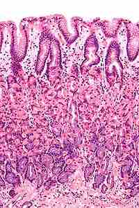
Like the other parts of the gastrointestinal tract, the stomach walls are made of a number of layers. From the inside to the outside, the first main layer is the mucosa. This consists of an epithelium, the lamina propria underneath, and a thin bit of smooth muscle called the muscularis mucosae. The submucosa lies under this and consists of fibrous connective tissue, separating the mucosa from the next layer, the muscularis externa. The muscularis in the stomach differs from that of other GI organs in that it has three layers of muscle instead of two. Under these muscle layers is the adventitia, layers of connective tissue continuous with the omenta.

The epithelium of the stomach forms deep pits, called fundic or oxyntic glands. Different types of cells are at different locations down the pits. The cells at the base of these pits are chief cells, responsible for production of pepsinogen, an inactive precursor of pepsin, which degrades proteins. The secretion of pepsinogen prevents self-digestion of the stomach cells. Further up the pits, parietal cells produce gastric acid and a vital substance, intrinsic factor. The function of gastric acid is two fold: 1) it kills most of the bacteria in food, stimulates hunger, and activates pepsinogen into pepsin; and 2) it denatures the complex protein molecule as a precursor to protein digestion through enzyme action in the stomach and small intestines. Near the top of the pits, closest to the contents of the stomach, there are mucous-producing cells called goblet cells that help protect the stomach from self-digestion.

The muscularis externa is made up of three layers of smooth muscle. The innermost layer is obliquely-oriented: this is not seen in other parts of the digestive system. This layer is responsible for creating the motion that churns and physically breaks down the food. The next layers are the square and then the longitudinal, which are present as in other parts of the GI tract. The pyloric antrum has thicker skin cells in its walls and performs more forceful contractions than the fundus. The pylorus is surrounded by a thick circular muscular wall which is normally tonically constricted, forming a functional (if not anatomically discrete) pyloric sphincter, which controls the movement of chyme.

The Stomach Wall

Micrograph showing a cross section of the stomach wall, in the body portion of the stomach. H&E stain.

[ edit ]
Edit this content
Prev Concept
Gross Anatomy of the Stomach
Digestive Properties of the Stomach
Next Concept
Subjects
  • Accounting
  • Algebra
  • Art History
  • Biology
  • Business
  • Calculus
  • Chemistry
  • Communications
  • Economics
  • Finance
  • Management
  • Marketing
  • Microbiology
  • Physics
  • Physiology
  • Political Science
  • Psychology
  • Sociology
  • Statistics
  • U.S. History
  • World History
  • Writing

Except where noted, content and user contributions on this site are licensed under CC BY-SA 4.0 with attribution required.