The stomach is a thick walled organ that lies between the esophagus and the first part of the small intestine (the duodenum). It is on the left side of the abdominal cavity, the fundus of the stomach lying against the diaphragm. Lying beneath the stomach is the pancreas. The greater omentum hangs from the greater curvature.
A mucous membrane lines the stomach which contains glands (with chief cells) that secrete gastric juices. Up to three quarts of this digestive fluid is produced daily. The gastric glands begin secreting before food enters the stomach due to the parasympathetic impulses of the vagus nerve, making the stomach also a storage vat for that acid.
The stomach is divided into four sections, each of which has different cells and functions . The sections are:
Sections of the Stomach
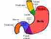
Diagram of the stomach, showing Cardiac region, Fundus, Body, and Pylorus.
- Cardiac region, where the contents of the esophagus empty into the stomach;
- Fundus, formed by the upper curvature of the organ;
- Body, the main central region;
- Pylorus or atrium, the lower section of the organ that facilitates emptying the contents into the small intestine.
Two smooth muscle valves, or sphincters, keep the contents of the stomach contained:
- Cardiac or esophageal sphincter, dividing the tract above;
- Pyloric sphincter or pyloric orifice, dividing the stomach from the small intestine.
The arteries supplying the stomach are the left gastric, the right gastric and right gastroepiploic branches of the hepatic, and the left gastroepiploic and short gastric branches of the lineal. They supply the muscular coat, ramify in the submucous coat, and are finally distributed to the mucous membrane.
The arteries break up at the base of the gastric tubules into a plexus of fine capillaries, which run upward between the tubules. They anatomize with each other and end in a plexus of larger capillaries, which surround the mouths of the tubes and also form hexagonal meshes around the ducts.
From these the veins arise, and pursue a straight course downward, between the tubules, to the submucous tissue; they end either in the lineal and superior mesenteric veins or directly in the portal vein.
The lymphatics are numerous. They consist of a superficial and a deep set, and pass to the lymph glands found along the two curvatures of the organ.
The nerves are the terminal branches of the right and left urethra and other parts, the former being distributed upon the back, and the latter upon the front part of the organ. A great number of branches from the celiac plexus of the sympathetic are also distributed to it. Nerve plexuses are found in the submucous coat and between the layers of the muscular coat as in the intestine. From these plexuses fibrils are distributed to the muscular tissue and the mucous membrane.