The development of the reproductive system is a part of prenatal development, and concerns the sex organs. Because its location to a large extent overlaps the urinary system, the development of them can also be described together as the development of the urinary and reproductive organs.
The reproductive organs are developed from the intermediate mesoderm. The permanent organs of the adult are preceded by a set of structures that are purely embryonic, and that with the exception of the ducts disappear almost entirely before the end of fetal life. These embryonic structures are the Wolffian and Müllerian ducts, also known as mesonephric and paramesonephric ducts, respectively. The Wolffian duct remains as the duct in males, and the Müllerian as that of the female.
In the male the Wolffian duct persists and forms the tube of the epididymis, the ductus deferens, and the ejaculatory duct, while the seminal vesicle arises during the third month as a lateral diverticulum from its hinder end. A large part of the head end of the mesonephros atrophies and disappears; of the remainder the anterior tubules form the efferent ducts of the testis; while the posterior tubules are represented by the ductuli aberrantes, and by the paradidymis, which is sometimes found in front of the spermatic cord above the head of the epididymis. In the female the Wolffian bodies and ducts atrophy.
Shortly after the formation of the Wolffian ducts a second pair of ducts is developed; these are the Müllerian ducts. In the male the Müllerian ducts atrophy, but in the female the Müllerian ducts persist and undergo further development. The portions that lie in the genital cord fuse to form the uterus and vagina. This fusion of the Müllerian ducts begins in the third month, and the septum formed by their fused medial walls disappears from below upward. The parts outside this cord remain separate and each forms the corresponding Fallopian tube.
About the fifth month a ring-like constriction marks the position of the cervix of the uterus, and after the sixth month the walls of the uterus begin to thicken. For a time the vagina is represented by a solid rod of epithelial cells. A ring-like outgrowth of this epithelium occurs at the lower end of the uterus and marks the future vaginal fornix. At about the fifth or sixth month the lumen of the vagina is produced by the breaking down of the central cells of the epithelium. The hymen represents the remains of the Müllerian eminence.
The gonads are the precursors of the testes in males and ovaries in females. They initially develop from the mesothelial layer of the peritoneum. The ovary is differentiated into a central part, the medulla of ovary, covered by a surface layer, the germinal epithelium. The immature ova originate from cells from the dorsal endoderm of the yolk sac.
The periphery of the testes is converted into the tunica albuginea. Cords of the central mass run together and form a network that becomes the rete testis, and another network, that develops the seminiferous tubules. In short, the descent of the testes consists of the opening of a connection from the testis to its final location at the anterior abdominal wall, followed by the development of the gubernaculum, which subsequently pulls and translocates the testis down into the developing scrotum. Ultimately, the passageway closes behind the testis. A failure in this process causes indirect inguinal hernia.
Until about the ninth week of gestational age, the external genitalia of males and females look the same, and follow a common development. This includes the development of a genital tubercle and a membrane dorsally to it, covering the developing urogenital opening, and the development of labioscrotal folds. Even after differentiation can be seen between the sexes, some stages are common, e.g. the disappearing of the membrane. On the other hand, sex-dependent development includes further protrusion of the genital tubercle in the male to form the penis. Furthermore, the labioscrotal folds evolve into the scrotum in males, while they evolve into labia in females.
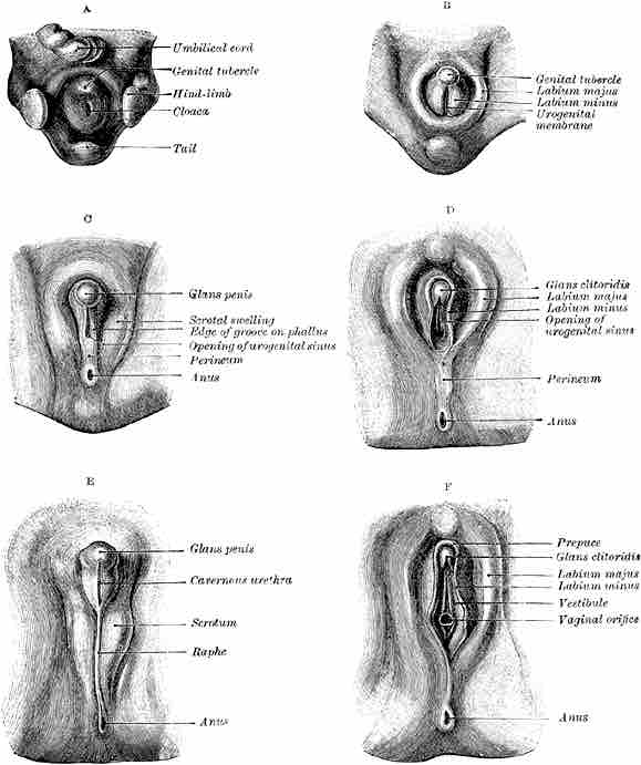
Development of male and female external genitalia from a common developmental beginning
A: common development. C,E: male development. B,D, F: female development.