Spinal Nerve Anatomy
The term spinal nerve generally refers to a mixed spinal nerve that carries motor, sensory, and autonomic signals between the spinal cord and the body.
Humans have 31 left–right pairs of spinal nerves, each roughly corresponding to a segment of the vertebral column: eight cervical spinal nerve pairs (C1–C8), 12 thoracic pairs (T1–T12), five lumbar pairs (L1–L5), five sacral pairs (S1–S5), and one coccygeal pair. The spinal nerves are part of the peripheral nervous system (PNS).

A spinal nerve
Spinal nerves arise from a combination of nerve fibers from the dorsal and ventral roots of the spinal cord.
LOCATION
Each spinal nerve is formed by the combination of nerve fibers from the dorsal and ventral roots of the spinal cord. The dorsal roots carry afferent sensory axons, while the ventral roots carry efferent motor axons.
The spinal nerve emerges from the spinal column through an opening (intervertebral foramen) between adjacent vertebrae.
Intervertebral foramina
Intervertebral foramina are indicated by arrows.
This is true for all spinal nerves except for the first spinal nerve pair, which emerges between the occipital bone and the atlas (the first vertebra). Thus the cervical nerves are numbered by the vertebra below, except C8, which exists below C7 and above T1.
The thoracic, lumbar, and sacral nerves are then numbered by the vertebra above. In the case of a lumbarized S1 vertebra (i.e., L6) or a sacralized L5 vertebra, the nerves are typically still counted to L5 and the next nerve is S1.
Spinal Nerve Innervation
Outside the vertebral column, the nerve divides into branches. The dorsal ramus contains nerves that serve the dorsal portions of the trunk; it carries visceral motor, somatic motor, and somatic sensory information to and from the skin and muscles of the back (epaxial muscles).
The ventral ramus contains nerves that serve the remaining ventral parts of the trunk and the upper and lower limbs (hypaxial muscles); they carry visceral motor, somatic motor, and sensory information to and from the ventrolateral body surface, structures in the body wall, and the limbs.
The meningeal branches (recurrent meningeal or sinuvertebral nerves) branch from the spinal nerve and re-enter the intervertebral foramen to serve the ligaments, dura, blood vessels, intervertebral discs, facet joints, and periosteum of the vertebrae.
The rami communicantes contain autonomic nerves that serve visceral functions, such as carrying visceral motor and sensory information to and from the visceral organs.
Cervical Nerves
The posterior distribution of the cervical nerves includes the suboccipital nerve (C1), the greater occipital nerve (C2), and the third occipital nerve (C3). The anterior distribution includes the cervical plexus (C1–C4) and brachial plexus (C5–T1).
The muscles innervated by the cervical nerves are the sternohyoid, sternothyroid, and omohyoid muscles.
A loop of nerves called ansa cervicalis is also part of the cervical plexus.
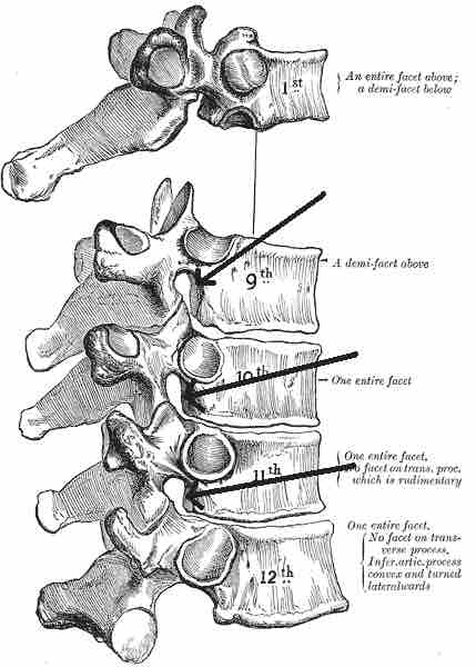
Thoracic Nerves
Thoracic nerve branches exit the spine and go directly to the paravertebral ganglia of the autonomic nervous system, where they are involved in the functions of organs and glands in the head, neck, thorax, and abdomen.
Anterior Divisions
The intercostal nerves come from thoracic nerves T1–T11, and run between the ribs. The subcostal nerve comes from nerve T12, and runs below the twelfth rib.
Posterior Divisions
The medial branches (ramus medialis) of the posterior branches of the upper six thoracic nerves run between the semispinalis dorsi and multifidus, which they supply.
They then pierce the rhomboid and trapezius muscles, and reach the skin by the sides of the spinous processes. This branch is called the medial cutaneous ramus.
The medial branches of the lower six thoracic nerves are distributed chiefly to the multifidus and longissimus dorsi, occasionally they give off filaments to the skin near the middle line. This sensitive branch is called the posterior cutaneous ramus.
Lumbar Nerves
The lumbar nerves are divided into posterior and anterior divisions.
Posterior Divisions
The medial branches of the posterior divisions of the lumbar nerves run close to the articular processes of the vertebrae and end in the multifidus muscle. The lateral branches supply the erector spinae muscles.
Anterior Divisions
The anterior divisions of the lumbar nerves (rami anteriores) consist of long, slender branches that accompany the lumbar arteries around the sides of the vertebral bodies, beneath the psoas major.
The first and second, and sometimes the third and fourth, lumbar nerves are each connected with the lumbar part of the sympathetic trunk by a white ramus communicans.
The nerves pass obliquely outward behind the psoas major, or between its fasciculi, distributing filaments to it and the quadratus lumborum.
The first three and the greater part of the fourth are connected by anastomotic loops and form the lumbar plexus.
The smaller part of the fourth joins with the fifth to form the lumbosacral trunk, which assists in the formation of the sacral plexus. The fourth nerve is named the furcal nerve, from the fact that it is subdivided between the two plexuses.
Sacral Nerves
There are five paired sacral nerves, half of them arising through the sacrum on the left side and the other half on the right side. Each nerve emerges in two divisions: one division through the anterior sacral foramina and the other division through the posterior sacral foramina.
The sacral nerves have both afferent and efferent fibers, thus they are responsible for part of the sensory perception and the movements of the lower extremities of the human body.
The pudendal nerve and parasympathetic fibers arise from S2, S3, and S4. They supply the descending colon and rectum, urinary bladder, and genital organs. These pathways have both afferent and efferent fibers.
Coccygeal Nerve
The coccygeal nerve is the 31st pair of spinal nerves and arises from the conus medullaris. Its anterior root helps form the coccygeal plexus.
Function
Spinal nerve motor functions are summarized in the table below.
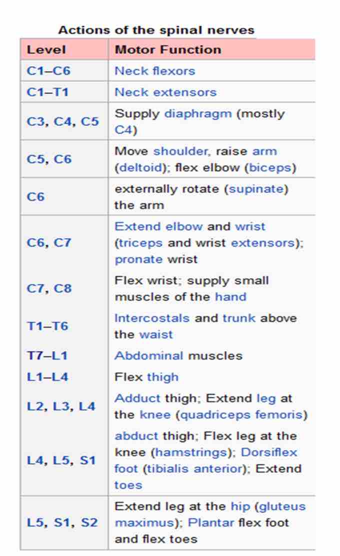
Spinal nerve function table
Spinal nerve motor functions are presented in a table.