Physiology
Textbooks
Boundless Anatomy and Physiology
Peripheral Nervous System
Cranial Nerves
Physiology Textbooks Boundless Anatomy and Physiology Peripheral Nervous System Cranial Nerves
Physiology Textbooks Boundless Anatomy and Physiology Peripheral Nervous System
Physiology Textbooks Boundless Anatomy and Physiology
Physiology Textbooks
Physiology
Concept Version 12
Created by Boundless

Oculomotor (III) Nerve

The oculomoter nerve (cranial nerve III) controls eye movement, such as constriction of the pupil and open eyelids.

Learning Objective

  • Describe the oculomotor nerve (cranial nerve III)


Key Points

    • The oculomotor nerve is the third paired cranial nerve.
    • The oculomotor nerve contains two nuclei, including the Edinger-Westphal nucleus that supplies parasympathetic nerve fibers to the eye to control pupil constriction and accommodation.
    • The oculomotor nerve originates at the superior colliculus and enters through the superior orbital fissure to control the levator palpebrae superioris muscles that hold the eyelids open.

Terms

  • tentorium cerebelli

    An extension of the dura mater that separates the cerebellum from the inferior portion of the occipital lobes.

  • cavernous sinus

    A venous channel found between layers of dura mater in the brain.

  • superior colliculus

    The primary integrating center for eye movements.

  • levator palpebrae superioris

    A muscle that elevates the upper eyelid.

  • plexus

    A network or interwoven mass, especially of nerves, blood vessels, or lymphatic vessels.

  • superior orbital fissure

    The superior orbital fissure is a foramen in the skull, although strictly it is more of a cleft, lying between the lesser and greater wings of the sphenoid bone.


Full Text

The oculomotor nerve is the third paired cranial nerve. It enters the orbit via the superior orbital fissure and controls most of the eye's movements, including constriction of the pupil and maintaining an open eyelid by innervating the levator palpebrae superiors muscle. 

The occulomotor nerve is derived from the basal plate of the embryonic midbrain. Cranial nerves IV and VI also participate in control of eye movement.

There are two nuclei for the oculomotor nerve:

  1. The oculomotor nucleus originates at the level of the superior colliculus. The muscles it controls are the striated muscle in the levator palpebrae superioris and all extraocular muscles, except for the superior oblique muscle and the lateral rectus muscle. 
  2. The Edinger-Westphal nucleus supplies parasympathetic fibers to the eye via the ciliary ganglion, and controls the pupillae muscle (affecting pupil constriction) and the ciliary muscle (affecting accommodation).

Sympathetic postganglionic fibers also join the nerve from the plexus on the internal carotid artery in the wall of the cavernous sinus and are distributed through the nerve, for example, to the smooth muscle of levator palpebrae superioris.

Emergence from Brain

On emerging from the brain, the oculomotor nerve is invested with a sheath of pia mater and enclosed in a prolongation from the arachnoid mater. It passes between the superior cerebellar and posterior cerebral arteries, and then pierces the dura mater anterior and lateral to the posterior clinoid process (to give attachment to the tectorium cerebella), passing between the free and attached borders of the tentorium cerebelli. 

It then runs along the lateral wall of the cavernous sinus, above the other orbital nerves, receiving in its course one or two filaments from the cavernous plexus of the sympathetic nervous system, and a communicating branch from the ophthalmic division of the trigeminal nerve.

It then divides into two branches that enter the orbit through the superior orbital fissure, between the two heads of the lateral rectus (a muscle on the lateral side of the eyeball in the orbit). Here the nerve is placed below the trochlear nerve and the frontal and lacrimal branches of the ophthalmic nerve, while the nasociliary nerve is placed between its two rami (the superior and inferior branch of oculomotor nerve).
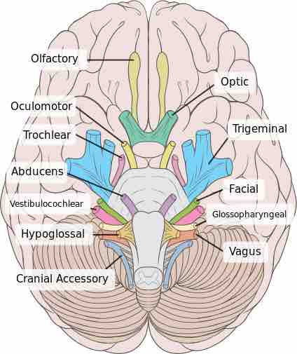
Cranial nerves

Image of cranial nerves showing the position of the oculomotor nerve.

This is an top view of the brain with the cranial nerves identified within it. The oculomotor nerve is the third nerve from the front of the brain, coming after the olfactory and optic nerves.
[ edit ]
Edit this content
Prev Concept
Optic (II) Nerve
Trochlear (IV) Nerve
Next Concept
Subjects
  • Accounting
  • Algebra
  • Art History
  • Biology
  • Business
  • Calculus
  • Chemistry
  • Communications
  • Economics
  • Finance
  • Management
  • Marketing
  • Microbiology
  • Physics
  • Physiology
  • Political Science
  • Psychology
  • Sociology
  • Statistics
  • U.S. History
  • World History
  • Writing

Except where noted, content and user contributions on this site are licensed under CC BY-SA 4.0 with attribution required.