Physiology
Textbooks
Boundless Anatomy and Physiology
Peripheral Nervous System
Cranial Nerves
Physiology Textbooks Boundless Anatomy and Physiology Peripheral Nervous System Cranial Nerves
Physiology Textbooks Boundless Anatomy and Physiology Peripheral Nervous System
Physiology Textbooks Boundless Anatomy and Physiology
Physiology Textbooks
Physiology
Concept Version 12
Created by Boundless

Abducens (VI) Nerve

The abducens nerve (cranial nerve VI) controls the lateral movement of the eye through innervation of the lateral rectus muscle.

Learning Objective

  • Describe the abducens nerve (cranial nerve VI)


Key Points

    • The abducens nerve exits the brainstem at the junction of the pons and the medulla and runs upward to reach the eye, traveling between the dura and the skull.
    • The long course of the abducens nerve between the brainstem and the eye makes it vulnerable to injury at many levels.
    • In most mammals besides humans, it also innervates the musculus retractor bulbi, which can retract the eye for protection.

Terms

  • lateral rectus muscle

    A muscle in the orbit. It is one of six extraocular muscles that control the movements of the eye (abduction in this case) and the only muscle innervated by the abducens nerve, cranial nerve VI, functioning to bring the pupil away from the midline of the body.

  • petrous temporal bone

    A pyramid-shaped bone that is wedged in at the base of the skull between the sphenoid and occipital bones and is part of the endocranium.

  • clivus

    A part of the cranium at the base of the skull. It forms a gradual sloping process at the anterior-most portion of the basilar occipital bone at its junction with the sphenoid bone.

  • abducens nerve

    A nerve that controls the lateral rectus muscle in the eye.


Full Text

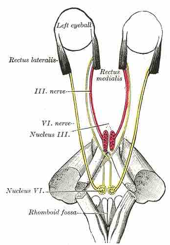
The abducens nerve (cranial nerve VI) is a somatic efferent nerve that, in humans, controls the movement of a single muscle: the lateral rectus muscle of the eye that moves the eye horizontally. In most other mammals it also innervates the musculus retractor bulbi, which can retract the eye for protection. Homologous abducens nerves are found in all vertebrates except lampreys and hagfishes.

The abducens nerve leaves the brainstem at the junction of the pons and the medulla, medial to the facial nerve. In order to reach the eye, it runs upward (superiorly) and then bends forward (anteriorly). 

The nerve enters the subarachnoid space when it emerges from the brainstem. It runs upward between the pons and the clivus, and then pierces the dura mater to run between the dura and the skull. 

At the tip of the petrous temporal bone, it makes a sharp turn forward to enter the cavernous sinus. In the cavernous sinus it runs alongside the internal carotid artery. It then enters the orbit through the superior orbital fissure and innervates the lateral rectus muscle of the eye.

Abducens nerve

Schematic of cranial nerves showing cranial nerve VI, the abducens nerve.

This is a schematic of the cranial nerves. In particular, it displays cranial nerve VI, the abducens nerve, and its connection from the eyes to the brainstem.

The long course of the abducens nerve between the brainstem and the eye makes it vulnerable to injury at many levels. For example, fractures of the petrous temporal bone can selectively damage the nerve, as can aneurysms of the intracavernous carotid artery. 

Mass lesions that push the brainstem downward can damage the nerve by stretching it between the point where it emerges from the pons and the point where it hooks over the petrous temporal bone.

[ edit ]
Edit this content
Prev Concept
Trigeminal (V) Nerve
Facial (VII) Nerve
Next Concept
Subjects
  • Accounting
  • Algebra
  • Art History
  • Biology
  • Business
  • Calculus
  • Chemistry
  • Communications
  • Economics
  • Finance
  • Management
  • Marketing
  • Microbiology
  • Physics
  • Physiology
  • Political Science
  • Psychology
  • Sociology
  • Statistics
  • U.S. History
  • World History
  • Writing

Except where noted, content and user contributions on this site are licensed under CC BY-SA 4.0 with attribution required.