The Dermis
Lying underneath the epidermis—the most superficial layer of our skin—is the dermis (sometimes called the corium). The dermis is a tough layer of skin. It is the layer of skin you touch when buying any leather goods.
The dermis is composed of two layers. They are the papillary layer (the upper layer) and the reticular layer (the lower layer).
The Papillary Layer
The papillary layer provides the layer above it, the epidermis, with nutrients to produce skin cells called keratinocytes. It also helps regulate the temperature of our skin and thus the body as a whole.
Both the nutrient supply and temperature regulation occur thanks to an extensive network of blood vessels in this layer. These blood vessels also help remove cellular waste products that would otherwise kill the skin cells if they were allowed to accumulate.
The pink tint to the skin of light-skinned individuals is due to the blood vessels found here. In fact, when you blush, it is the dilation of these blood vessels that causes you to turn red. The uneven projections found in this layer, called dermal papillae, also form people's fingerprints and give this layer its name.
The Reticular Layer
The reticular layer serves to strengthen the skin and also provides our skin with elasticity. Elasticity refers to how our skin is able to spring back into shape if deformed by something like a pinch. The reticular layer also contains hair follicles, sweat glands, and sebaceous glands.
The sweat gland can either be apocrine, such as those found in the armpits and the groin area, or the eccrine glands, which are found all over the body. The former help contribute to body odor (along with the bacteria on our skin), and the latter help regulate our body temperature through the process of evaporation.
The sebaceous glands found in the dermis secrete a substance called sebum that helps to lubricate and protect our skin from drying out.
The dermis also contains:
- Nerve endings that transmit various stimuli such as pain, itch, pressure, and temperature.
- Lymphatic vessels that transport immune system cells, the cells that help destroy infectious organisms that may have found their way into our body via a scratch on the skin.
- Collagen, a protein that helps strengthen our skin, and elastin, a protein that helps keep our skin flexible.
The Hypodermis
Beneath the dermis is the deepest layer of our skin. It is alternatively termed hypodermis, subcutis, or subcutaneous tissue. It contains many collagen cells as well as fat.
Fat, in particular, helps insulate our body from the cold and act as a cushion for our internal structures (such as muscles and organs) when something hits us. Fat can also be called upon by the body in times of great need as an energy source.
Given the alternative names for this layer, it should come as no surprise that this is the layer where subcutaneous injections are given into via a hypodermic needle.
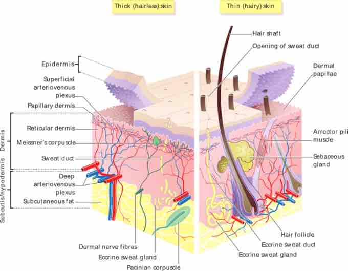
Skin sensory receptors
Those nearest the surface of the skin include receptors that detect gentle pressure, temperature, and vibrations, as well as naked nerve endings (dendrites) that detect pain. Deeper in the dermis are naked dendrites that wind around the bases of hair follicles and detect motions of the hairs, as well as receptors such as Pacinian corpuscles that respond to strong pressure and vibrations.
Human Skin
This image details the parts of the integumentary system.