Physiology
Textbooks
Boundless Anatomy and Physiology
Human Development and Pregnancy
Third Week of Development
Physiology Textbooks Boundless Anatomy and Physiology Human Development and Pregnancy Third Week of Development
Physiology Textbooks Boundless Anatomy and Physiology Human Development and Pregnancy
Physiology Textbooks Boundless Anatomy and Physiology
Physiology Textbooks
Physiology
Concept Version 13
Created by Boundless

Chorionic Villi and Placental Development

In the placenta, chorionic villi develop to maximize surface-area contact with the maternal blood for nutrient and gas exchange.

Learning Objective

  • Summarize the development of the chorionic villi and placenta


Key Points

    • Chorionic villi invade and destroy the uterine decidua while at the same time they absorb nutritive materials from it to support the growth of the embryo.
    • The villi begin primary development in the fourth week, becoming fully vascularized between the fifth and sixth weeks.
    • Placental development begins with implantation of the blastocyst; this leads to its differentiation into several layers that allow nutrient, gas, and waste exchange to the developing embryo and fetus—as well as forming a protective barrier.

Terms

  • chorion

    The protective and nutritive membrane that attaches higher vertebrate fetuses to the uterus.

  • uterine decidua

    The term for the uterine lining (endometrium) during a pregnancy, which forms the maternal part of the placenta. It is formed under the influence of progesterone and forms highly characteristic cells.

  • chorionic villi

    These sprout from the chorion in order to give a maximum area of contact with the maternal blood.

  • placenta

    A vascular organ present only in the female during gestation. It supplies food and oxygen from the mother to the fetus, and passes back waste. It is implanted in the wall of the uterus and links to the fetus through the umbilical cord. It is expelled after birth.


Full Text

Chorionic Villi

Chorionic villi sprout from the chorion after their rapid proliferation in order to give a maximum area of contact with the maternal blood. These villi invade and destroy the uterine decidua while at the same time they absorb nutritive materials from it to support the growth of the embryo .
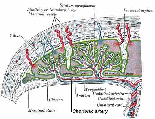
Chorionic artery

An image showing the chorionic villi and the maternal vessels.

This is a schematic drawing of a chorionic artery. It shows the chorionic villi connecting to the maternal vessels.

During the primary stage (the end of fourth week), the chorionic villi are small, nonvascular, and contain only the trophoblast. During the secondary stage (the fifth week), the villi increase in size and ramify, while the mesoderm grows into them; at this point the villi contain trophoblast and mesoderm. 

During the tertiary stage (fifth to sixth week), the branches of the umbilical vessels grow into the mesoderm; in this way, the chorionic villi are vascularized. At this point, the villi contain trophoblast, mesoderm, and blood vessels.

Embryonic blood is carried to the villi by the branches of the umbilical arteries. After circulating through the capillaries of the villi, it is returned to the embryo by the umbilical veins. Chorionic villi are vital in pregnancy from a histomorphologic perspective and are, by definition, products of conception.

Placenta

The placenta is a fetally derived organ that connects the developing fetus to the uterine wall to allow nutrient uptake, waste elimination, and gas exchange via the mother's blood supply. The placenta begins to develop upon implantation of the blastocyst into the maternal endometrium. 

The placenta functions as a fetomaternal organ with two components: the fetal placenta (chorion frondosum), which develops from the same blastocyst that forms the fetus; and the maternal placenta (decidua basalis), which develops from the maternal uterine tissue.

The outer layer of the blastocyst becomes the trophoblast, which forms the outer layer of the placenta. This layer is divided into two further layers: the underlying cytotrophoblast layer and the overlying syncytiotrophoblast layer. 

The latter is a multinucleated, continuous cell layer that covers the surface of the placenta. It forms as a result of the differentiation and fusion of the underlying cytotrophoblast cells, a process that continues throughout placental development. The syncytiotrophoblast (otherwise known as syncytium) thereby contributes to the barrier function of the placenta.
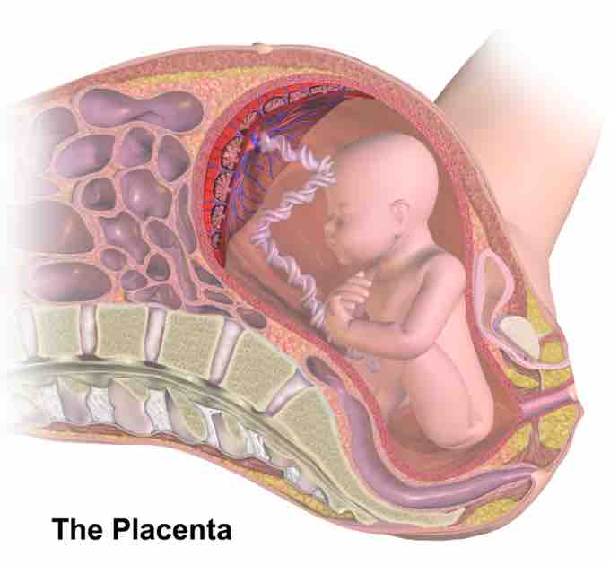
Placenta

Image illustrating the placenta and chorionic villi. The umbilical cord is seen connected to the fetus and the placenta.

This is a color image illustrating the placenta and chorionic villi. The umbilical cord is seen connecting the fetus to the placenta.
[ edit ]
Edit this content
Prev Concept
Development of the Cardiovascular System
Fourth Week of Development
Next Concept
Subjects
  • Accounting
  • Algebra
  • Art History
  • Biology
  • Business
  • Calculus
  • Chemistry
  • Communications
  • Economics
  • Finance
  • Management
  • Marketing
  • Microbiology
  • Physics
  • Physiology
  • Political Science
  • Psychology
  • Sociology
  • Statistics
  • U.S. History
  • World History
  • Writing

Except where noted, content and user contributions on this site are licensed under CC BY-SA 4.0 with attribution required.