The peritoneum is the serous membrane that forms the lining of the abdominal cavity or the coelom. It covers most of the intra-abdominal, or coelomic, organs. It is composed of a layer of mesothelial tissue, supported by a thin layer of connective tissue. The peritoneum provides support and protection for the abdominal organs, and is the main conduit for the associated lymph vessels, nerves, and abdominal arteries and veins.
The abdominal cavity is the open space surrounded by the vertebrae, abdominal muscles, diaphragm, and pelvic floor. Remember not to confuse the abdominal cavity with the intraperitoneal space, which is in fact located within the abdominal cavity, and wrapped in peritoneum tissue. For example, a kidney is inside the abdominal cavity, but is retroperitoneal - located outside the peritoneum .
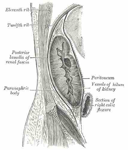
The Peritoneum and the Kidney
Sagittal section through posterior abdominal wall, showing the kidney residing outside the peritoneum.
Although they ultimately form one continuous sheet, there are two layers of peritoneum and potential space between those layers. The outer layer, called the parietal peritoneum, is attached to the abdominal wall. The inner layer, the visceral peritoneum, is wrapped around the internal organs that are located inside the intraperitoneal cavity. The potential space between these two layers is the peritoneal cavity. It is filled with a small amount of slippery serous fluid that allows the two layers to slide freely over each other.
The term "mesentery" is often used to refer to a double layer of visceral peritoneum. There are generally blood vessels, nerves, and other structures between these layers. This space between the two layers is technically outside of the peritoneal sac, and thus not in the peritoneal cavity.
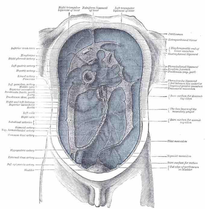
There are two main regions of the peritoneum, connected by the epiploic foramen (also known as the omental foramen or foramen of Winslow). The first is the greater sac or general cavity of the abdomen, and is represented in red in the image below. The second is the lesser sac or omental bursa, and is represented in blue in the image. The lesser sac is divided into two "omenta": the gastrohepatic and the gastrocolic. The gastrohepatic omentum is attached to the lesser curvature of the stomach and the liver. The gastrocolic omentum hangs from the greater curve of the stomach and loops down in front of the intestines before curving back upwards to attach to the transverse colon. Like a curtain of tissue, it is draped in front of the intestines, serving to insulate and protect.

Substructures of the Peritoneum
The epiploic foramen, greater sac or general cavity (red) and lesser sac, or omental bursa (blue). Midsagittal cross-section.
The structures in the abdomen are classified as intraperitoneal, retroperitoneal, or infraperitoneal depending on whether they are covered with visceral peritoneum and whether they are attached by mesenteries, such as the mensentery and mesocolon. Intraperitoneal organs include the stomach, the first five centimeters and the fourth part of the the duodenum, the jejunum, the ileum, the cecum, the appendix, the transverse colon, the sigmoid colon, and the upper third of the rectum. Other organs located in the intraperitoneal space are the liver, spleen, and the tail of the pancreas. In women, the uterus, fallopian tubes, ovaries, and gonadal blood vessels are located in the intraperitoneum.
Retroperitoneal structures include the rest of the duodenum, the ascending colon, the descending colon, the middle third of the rectum, and the remainder of the pancreas. Other organs located in the retroperitoneal space are the kidneys, adrenal glands, proximal ureters, and renal vessels. Organs located below the peritoneum in the subperitoneal space include the lower third of the rectum and the urinary bladder.
Structures that are intraperitoneal are generally mobile, while those that are retroperitoneal are relatively fixed in their location. Some structures, such as the kidneys, are "primarily retroperitoneal," while others such as the majority of the duodenum, are "secondarily retroperitoneal", meaning that structure developed intraperitoneally, but lost its mesentery and thus became retroperitoneal.
Peritoneum
The peritoneum illustrated, indicated by blue.