Physiology
Textbooks
Boundless Anatomy and Physiology
Cardiovascular System: Blood Vessels
The Venous System
Physiology Textbooks Boundless Anatomy and Physiology Cardiovascular System: Blood Vessels The Venous System
Physiology Textbooks Boundless Anatomy and Physiology Cardiovascular System: Blood Vessels
Physiology Textbooks Boundless Anatomy and Physiology
Physiology Textbooks
Physiology
Concept Version 12
Created by Boundless

Venules

Venules are small blood vessels in the microcirculation that connect capillary beds to veins.

Learning Objective

  • Describe the venules of the venous system


Key Points

    • Many venules unite to form a vein.
    • Venule walls have three layers: an inner endothelium composed of squamous endothelial cells that act as a membrane, a middle layer of muscle and elastic tissue, and an outer layer of fibrous connective tissue.
    • High-endothelial venules are specialized post-capillary venous swellings characterized by plump endothelial cells, in contrast with the thinner endothelial cells found in regular venules.

Terms

  • venule

    A small blood vessel in the microcirculation that allows deoxygenated blood to return from capillary beds to veins.

  • high endothelial venule

    A specialized post-capillary venous swelling of the lymphatic system that allows lymphocytes (white blood cells) to easily exit the circulatory system.


Full Text

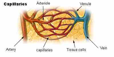
A venule is a small blood vessel in the microcirculation that allows deoxygenated blood to return from capillary beds to larger blood vessels called veins. Venules range from 8 to 100μm in diameter and are formed when capillaries come together. Many venules unite to form a vein.

Venule walls have three layers: an inner endothelium composed of squamous endothelial cells that act as a membrane, a middle layer of muscle and elastic tissue, and an outer layer of fibrous connective tissue. The middle layer is poorly developed so that venules have thinner walls than arterioles. Venules are extremely porous so that fluid and blood cells can move easily from the bloodstream through their walls.

Venule

Venules form when capillaries come together and converging venules form a vein.

This diagram indicates capillaries, arteries, arterioles, venules, tissue cells, and veins.

In contrast to regular venules, high-endothelial venules (HEV) are specialized post-capillary venous swellings. They are characterized by plump endothelial cells as opposed to the usual thinner endothelial cells found in regular venules. HEVs enable lymphocytes (white blood cells) circulating in the blood to directly enter a lymph node by crossing through the HEV.

[ edit ]
Edit this content
Prev Concept
Capillaries
Veins
Next Concept
Subjects
  • Accounting
  • Algebra
  • Art History
  • Biology
  • Business
  • Calculus
  • Chemistry
  • Communications
  • Economics
  • Finance
  • Management
  • Marketing
  • Microbiology
  • Physics
  • Physiology
  • Political Science
  • Psychology
  • Sociology
  • Statistics
  • U.S. History
  • World History
  • Writing

Except where noted, content and user contributions on this site are licensed under CC BY-SA 4.0 with attribution required.