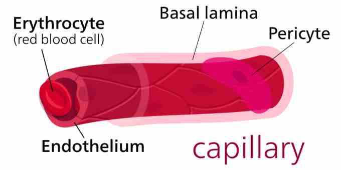
Capillaries, which form part of the micro-circulation, are the smallest of the body's blood vessels at between 5-10 μm in diameter with the endothelial vessel wall of only one cell thick. They are surrounded by a thin basal lamina of connective tissue.
Structure of a capillary
Capillaries are of small diameter with the vessel wall being a single cell thick. Capillaries are surrounded by a thin basal lamina of connective tissue.
Capillary Function
Capillaries form a network through body tissues that connects arterioles and venules and facilitates the exchange of water, oxygen, carbon dioxide, and many other nutrients and waste substances between blood and surrounding tissues. The thin wall of the capillary and close association with its resident tissue allow for gas and lipophilic molecules to pass through without the need for special transport mechanisms. This allows bidirectional diffusion depending on osmotic gradients.
Formation of New Capillaries
During embryological development, new capillaries are formed by vasculogenesis, the process of blood vessel formation occurring by de novo production of endothelial cells and their formation into vascular tubes. The term angiogenesis denotes the formation of new capillaries from pre-existing blood vessels.
The Capillary Bed
Capillaries do not function independently. The capillary bed is an interwoven network of capillaries that supplies an organ. The more metabolically active the cells, the more capillaries required to supply nutrients and carry away waste products.
A capillary bed can consist of two types of vessels: true capillaries, which branch mainly from arterioles and provide exchange between cells and the circulation, and vascular shunts, short vessels that directly connect arterioles and venules at opposite ends of the bed, allowing for bypass.
Types of Capillaries
There are three main types of capillaries:
- Continuous - Endothelial cells provide an uninterrupted lining, only allowing small molecules like water and ions to diffuse through tight junctions. This leave gaps of unjoined membrane called intercellular clefts.
- Fenestrated - Fenestrated capillaries have pores in the endothelial cells (60-80 nanometers in diameter) that are spanned by a diaphragm of radially-oriented fibrils. They allow small molecules and limited amounts of protein to diffuse.
- Sinusoidal - Sinusoidal capillaries are a special type of fenestrated capillaries that have larger openings (30-40 μm in diameter) in the endothelium. These types of blood vessels allow red and white blood cells (7.5μm - 25μm diameter) and various serum proteins to pass using a process aided by a discontinuous basal lamina. Sinusoid blood vessels are primarily located in the bone marrow, lymph nodes, and adrenal gland. Some sinusoids are special in that they do not have tight junctions between cells. These are called discontinuous sinusoidal capillaries, present in the liver and spleen where greater movement of cells and materials is necessary.
Types of Capillary
The three types of capillaries: continuous, fenestrated and sinusoid.
Control of Flow
Capillary beds may control blood flow via autoregulation. This allows an organ to maintain constant flow despite a change in central blood pressure. This is achieved by myogenic response and by tubuloglomerular feedback in the kidney. When blood pressure increases, the arterioles that lead to the capillary bed are stretched and subsequently constrict to counteract the increased tendency for high pressure to increase blood flow. In the lungs, special mechanisms have been adapted to meet the needs of increased necessity of blood flow during exercise. When heart rate increases and more blood must flow through the lungs, capillaries are recruited and are distended to make room for increased blood flow while resistance decreases.