Physiology
Textbooks
Boundless Anatomy and Physiology
Cardiovascular System: Blood Vessels
Arteries
Physiology Textbooks Boundless Anatomy and Physiology Cardiovascular System: Blood Vessels Arteries
Physiology Textbooks Boundless Anatomy and Physiology Cardiovascular System: Blood Vessels
Physiology Textbooks Boundless Anatomy and Physiology
Physiology Textbooks
Physiology
Concept Version 14
Created by Boundless

Elastic Arteries

An elastic or conducting artery has a large number of collagen and elastin filaments in the tunica media.

Learning Objective

  • Distinguish the elastic artery from the muscular artery


Key Points

    • Elastic arteries include the largest arteries in the body, those closest to the heart. They give rise to medium-sized vessels known as muscular, or distributing, arteries.
    • Elastic arteries differ from muscular arteries both in size and in the relative amount of elastic tissue contained within the tunica media.
    • Arterial elasticity gives rise to the Windkessel effect, which helps to maintain a relatively constant pressure in the arteries despite the pulsating nature of blood flow.

Terms

  • elastic arteries

    An artery with a large number of collagen and elastin filaments, giving it the ability to stretch in response to each pulse.

  • tunica media

    The middle layer of a vein wall with bands of thin smooth muscle.


Full Text

Elastic arteries contain larger numbers of collagen and elastin filaments in their tunica media than muscular arteries do, giving them the ability to stretch in response to each pulse.

Elastic arteries include the largest arteries in the body, those closest to the heart, and give rise to the smaller muscular arteries. The pulmonary arteries, the aorta, and its branches together comprise the body's system of elastic arteries. In these large arteries, the amount of elastic tissue is considerable and the smooth muscle fiber cells are arranged in 5 to 7 layers in both circular and longitudinal directions.

Arterial elasticity gives rise to the Windkessel effect, which through passive contraction after expansion helps to maintain a relatively constant pressure in the arteries despite the pulsating nature of the blood flow from the heart.

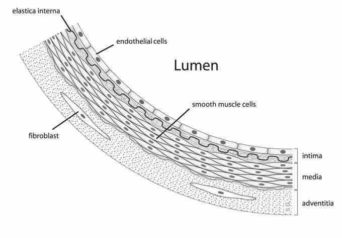
Anatomy of the Arterial Wall

Arterial wall layers including the tunica intima and the tunica media. In elastic arteries, the tunica media is rich with elastic and connective tissue.
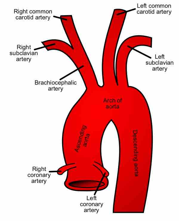
This diagram of the arterial wall indicates the fibroblast, elastica interna, endothelial cells, lumen, smooth muscle cells, intima, media, and adventitia.

The Aorta

Due to position as the first part of the systemic circulatory system closest to the heart and the resultant high pressures it will experience, the aorta is perhaps the most elastic artery, featuring an incredibly thick tunica media rich in elastic filaments. The aorta is so thick that it requires its own capillary network to supply it with sufficient oxygen and nutrients to function, the vasa vasorum.

When the left ventricle contracts to force blood into the aorta, the aorta expands. This stretching generates the potential energy that will help maintain blood pressure during diastole, when the aorta contracts passively. Additionally, the elastic recoil helps conserve the energy from the pumping heart and smooth the flow of blood around the body through the Windkessel effect.

The aorta

The aorta makes up most of the elastic arteries in the body.

This diagram of the aorta indicates the left and right common carotid arteries, left and right subclavian arteries, brachiocephalic artery, aortic arch, ascending and descending aorta, and right and left coronary arteries.
[ edit ]
Edit this content
Prev Concept
Artery Function
Muscular Arteries
Next Concept
Subjects
  • Accounting
  • Algebra
  • Art History
  • Biology
  • Business
  • Calculus
  • Chemistry
  • Communications
  • Economics
  • Finance
  • Management
  • Marketing
  • Microbiology
  • Physics
  • Physiology
  • Political Science
  • Psychology
  • Sociology
  • Statistics
  • U.S. History
  • World History
  • Writing

Except where noted, content and user contributions on this site are licensed under CC BY-SA 4.0 with attribution required.