Physiology
Textbooks
Boundless Anatomy and Physiology
APPENDIX B: Development and Aging of the Organ Systems
Integumentary System Development
Physiology Textbooks Boundless Anatomy and Physiology APPENDIX B: Development and Aging of the Organ Systems Integumentary System Development
Physiology Textbooks Boundless Anatomy and Physiology APPENDIX B: Development and Aging of the Organ Systems
Physiology Textbooks Boundless Anatomy and Physiology
Physiology Textbooks
Physiology
Concept Version 13
Created by Boundless

Development of the Integumentary System

The integumentary system develops from all embryonic layers (ectoderm, mesoderm and neural crest cells).

Learning Objective

  • Outline the developmental process of the integumentary system


Key Points

    • Ectoderm forms at 4 weeks.
    • Between 4 and 12 weeks, the stratified epithelium forms and mesoderm forms the blood vessels and connective tissue.
    • At 16 weeks, the basement membrane folds and melanoblasts start producing melanin.
    • At 20 weeks, hair begins to grow from sebaceous glands and sweat glands form.

Terms

  • fetal skin

    Fetal skin forms from three layers: ectoderm, mesoderm, and neural crest cells.

  • neural crest cells

    Neural crest cells are a transient, multipotent, migratory cell population that gives rise to a diverse cell lineage including melanocytes, craniofacial cartilage and bone, smooth muscle, peripheral and enteric neurons, and glia.

  • sebaceous gland

    A gland of the skin that secretes an oily substance, sebum, usually into a hair follicle near the surface of the skin.


Full Text

OVERVIEW

The integumentary system is the largest organ system in the human body, and is responsible for protecting the body from most physical and environmental factors. The largest organ in the body is the skin. The integumentary system is both a barrier and a sensory organ. The integument also includes appendages, primarily the sweat and sebaceous glands, hair, nails and arrectores pillorum (tiny muscles at the root of each hair that cause goose bumps).

FORMATION OF SKIN DURING GESTATION

Fetal skin forms from three layers: ectoderm, mesoderm, and neural crest cells. Figure 1 shows a diagram of the skin structure.

At four weeks gestation, simple ectoderm epithelium forms. Between four and 12 weeks, the basal cellls divide repeatedly forming the stratified epithelium. During this same period, the mesoderm forms the blood vessels and connective tissues. Epidermal ridges (e.g. fingerprints) begin to develop around 10 weeks gestation and are completed by 17 weeks gestation. Sensory nerves also develop.

At 16 weeks gestation, the basement membrane folds. Melanoblasts that will form melanocytes migrate with neural crests cells to the epithelium and begin producing melanin prior to birth. The connective tissue differentiates into the various layers of the dermis. Ectoderm thickens into finger and toe nails. Other regions of the ectoderm form into epithelial columns called cords which will become hair follocles and sebaceous and sweat glands.

At 20 weeks gestation, hair begins to grow from sebaceous glands, while sweat glands are formed from coiled cords. Other cords begin to form mammary glands.
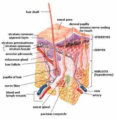
Human Skin

This image details the parts of the integumentary system.

[ edit ]
Edit this content
Prev Concept
Infertility
Effects of Aging on the Integumentary System
Next Concept
Subjects
  • Accounting
  • Algebra
  • Art History
  • Biology
  • Business
  • Calculus
  • Chemistry
  • Communications
  • Economics
  • Finance
  • Management
  • Marketing
  • Microbiology
  • Physics
  • Physiology
  • Political Science
  • Psychology
  • Sociology
  • Statistics
  • U.S. History
  • World History
  • Writing

Except where noted, content and user contributions on this site are licensed under CC BY-SA 4.0 with attribution required.