Physiology
Textbooks
Boundless Anatomy and Physiology
APPENDIX B: Development and Aging of the Organ Systems
Development of Metabolism
Physiology Textbooks Boundless Anatomy and Physiology APPENDIX B: Development and Aging of the Organ Systems Development of Metabolism
Physiology Textbooks Boundless Anatomy and Physiology APPENDIX B: Development and Aging of the Organ Systems
Physiology Textbooks Boundless Anatomy and Physiology
Physiology Textbooks
Physiology
Concept Version 12
Created by Boundless

Development of Metabolism

The gut or gastrointestinal tract is an endoderm-derived structure.

Learning Objective

  • Describe changes in metabolism associated with aging


Key Points

    • At approximately the sixteenth day of human development, the embryo begins to fold ventrally in two directions. As a result, a piece of the yolk sac is pinched off to become the primitive gut. The yolk sac remains connected to the gut tube via the vitelline duct.
    • During fetal life, the primitive gut can be divided into three segments: foregut, midgut, and hindgut. In later development each segment of the gut gives rise to specific gut and gut-related structures.
    • Components derived from the gut proper, including the stomach and colon, develop as swellings or dilatations of the primitive gut.
    • Gut-related derivatives develop as out-pouchings of the primitive gut.
    • The blood vessels supplying these structures remain constant throughout development.

Terms

  • foregut

    The anterior part of the alimentary canal of an embryo, from the mouth to the duodenum.

  • midgut

    The midgut is the portion of the embryo from which most of the intestines develop.

  • hindgut

    The hindgut (or epigaster) is the posterior (caudal) part of the alimentary canal. In mammals, it includes the distal third of the transverse colon and the splenic flexure, the descending colon, sigmoid colon, and rectum.


Full Text

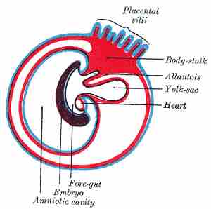
The gut or gastrointestinal tract is an endoderm-derived structure. At approximately the sixteenth day of human development, the embryo begins to fold ventrally (with the embryo's ventral surface becoming concave) in two directions: the sides of the embryo fold in on each other and the head and tail fold toward one another.

As a result, a piece of the yolk sac (the endoderm-lined structure in contact with the ventral aspect of the embryo) is then "pinched off" to become the primitive gut. The yolk sac remains connected to the gut tube via the vitelline duct. Usually this structure regresses during development; in cases where it does not, it is known as Meckel's diverticulum.

During fetal life, the primitive gut can be divided into three segments: foregut , midgut , and hindgut . These terms are regularly used to describe both segments of the primitive gut and components of the definitive gut.
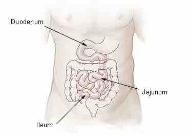
Small Intestine

The midgut is the portion of the embryo from which most of the intestines develop. The hindgut (or epigaster) is the posterior (caudal) part of the alimentary canal.

In later development each segment of the gut gives rise to specific gut and gut-related structures. Components derived from the gut proper, including the stomach and colon, develop as swellings or dilatations of the primitive gut. In contrast, gut-related derivatives—that is, those structures that derive from the primitive gut but are not part of the gut proper—mainly develop as out-pouchings of the primitive gut. The blood vessels supplying these structures remain constant throughout development.

Foregut

Diagram showing the expansion of amnion and delimitation of the umbilicus.

[ edit ]
Edit this content
Prev Concept
Aging and the Digestive System
Development of the Urinary System
Next Concept
Subjects
  • Accounting
  • Algebra
  • Art History
  • Biology
  • Business
  • Calculus
  • Chemistry
  • Communications
  • Economics
  • Finance
  • Management
  • Marketing
  • Microbiology
  • Physics
  • Physiology
  • Political Science
  • Psychology
  • Sociology
  • Statistics
  • U.S. History
  • World History
  • Writing

Except where noted, content and user contributions on this site are licensed under CC BY-SA 4.0 with attribution required.