Physiology
Textbooks
Boundless Anatomy and Physiology
APPENDIX B: Development and Aging of the Organ Systems
Development of Lymphatic Tissues
Physiology Textbooks Boundless Anatomy and Physiology APPENDIX B: Development and Aging of the Organ Systems Development of Lymphatic Tissues
Physiology Textbooks Boundless Anatomy and Physiology APPENDIX B: Development and Aging of the Organ Systems
Physiology Textbooks Boundless Anatomy and Physiology
Physiology Textbooks
Physiology
Concept Version 13
Created by Boundless

Lymphatic Tissue Development

Lymphatic tissue development begins by the end of the fifth week of embryonic development.

Learning Objective

  • Describe lymphatic tissue development


Key Points

    • The first lymph sacs to appear are the paired jugular lymph sacs at the junction of the internal jugular and subclavian veins.
    • The next lymph sac to appear is the unpaired retroperitoneal lymph sac at the root of the mesentery of the intestine. It develops from the primitive vena cava and mesonephric veins.
    • The last of the lymph sacs, the paired posterior lymph sacs, develop from the iliac veins. The posterior lymph sacs produce capillary plexuses and lymphatic vessels of the abdominal wall, pelvic region, and lower limbs.
    • With the exception of the anterior part of the sac from which the cisterna chyli develops, all lymph sacs become invaded by mesenchymal cells and are converted into groups of lymph nodes.
    • The spleen develops from mesenchymal cells between layers of the dorsal mesentery of the stomach.

Terms

  • cisterna chyli

    a dilated sac at the lower end of the thoracic duct into which lymph from the intestinal trunk and two lumbar lymphatic trunks flows

  • lymph node

    small oval bodies of the lymphatic system, distributed along the lymphatic vessels, that are clustered in the armpits, groin, neck, chest, and abdomen. They act as filters, with an internal honeycomb of connective tissue filled with lymphocytes and macrophages that collect and destroy bacteria, viruses, and foreign matter from lymph. When the body is fighting an infection, these lymphocytes multiply rapidly and produce a characteristic swelling of the lymph nodes.

  • mesoderm

    One of the three tissue layers in the embryo of a metazoan animal. Through embryonic development, it will produce many internal organs of the adult, e.g. muscles, spine, and circulatory system.

  • lymph sac

    The lymph sacs are precursors of the lymph vessels.


Full Text

Lymphatic tissues begin to develop by the end of the fifth week of embryonic development. Lymphatic vessels develop from lymph sacs that arise from developing veins, which are derived from mesoderm.

The first lymph sacs to appear are the paired jugular lymph sacs at the junction of the internal jugular and subclavian veins. From the jugular lymph sacs, lymphatic capillary plexuses spread to the thorax, upper limbs, neck, and head. Some of the plexuses enlarge and form lymphatic vessels in their respective regions. Each jugular lymph sac retains at least one connection with its jugular vein, the left one developing into the superior portion of the thoracic duct. The next lymph sac to appear is the unpaired retroperitoneal lymph sac at the root of the mesentery of the intestine. It develops from the primitive vena cava and mesonephric veins. Capillary plexuses and lymphatic vessels spread from the retroperitoneal lymph sac to the abdominal viscera and diaphragm. The sac establishes connections with the cisterna chyli, but loses its connections with neighboring veins.

The last of the lymph sacs, the paired posterior lymph sacs, develop from the iliac veins. The posterior lymph sacs produce capillary plexuses and lymphatic vessels of the abdominal wall, pelvic region, and lower limbs. The posterior lymph sacs join the cisterna chyli and lose their connections with adjacent veins. With the exception of the anterior part of the sac from which the cisterna chyli develops, all lymph sacs become invaded by mesenchymal cells and are converted into groups of lymph nodes . The spleen develops from mesenchymal cells between layers of the dorsal mesentery of the stomach. The thymus arises as an outgrowth of the third pharyngeal pouch.

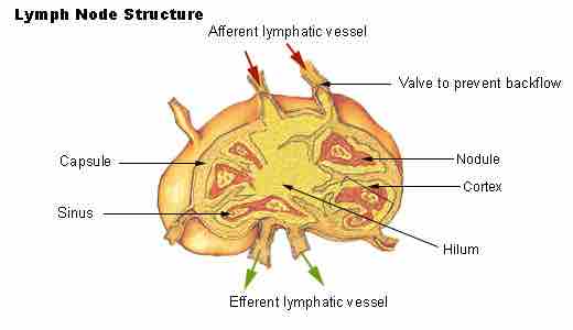
Lymph node structure

A lymph node showing afferent and efferent lymphatic vessels.

[ edit ]
Edit this content
Prev Concept
Aging and the Cardiovascular System
Development of the Immune System
Next Concept
Subjects
  • Accounting
  • Algebra
  • Art History
  • Biology
  • Business
  • Calculus
  • Chemistry
  • Communications
  • Economics
  • Finance
  • Management
  • Marketing
  • Microbiology
  • Physics
  • Physiology
  • Political Science
  • Psychology
  • Sociology
  • Statistics
  • U.S. History
  • World History
  • Writing

Except where noted, content and user contributions on this site are licensed under CC BY-SA 4.0 with attribution required.