The human eye is the gateway to one of our five senses. The human eye is an organ that reacts with light. It allows light perception, color vision and depth perception. A normal human eye can see about 10 million different colors! There are many parts of a human eye, and that is what we are going to cover in this atom.
Properties
Contrary to what you might think, the human eye is not a perfect sphere, but is made up of two differently shaped pieces, the cornea and the sclera. These two parts are connected by a ring called the limbus. The part of the eye that is seen is the iris, which is the colorful part of the eye. In the middle of the iris is the pupil, the black dot that changes size. The cornea covers these elements, but is transparent. The fundus is on the opposite of the pupil, but inside the eye and can not be seen without special instruments. The optic nerve is what conveys the signals of the eye to the brain. is a diagram of the eye. The human eye is made up of three coats:
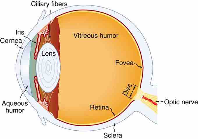
Diagram of the Human Eye
The cornea and lens of an eye act together to form a real image on the light-sensing retina, which has its densest concentration of receptors in the fovea and a blind spot over the optic nerve. The power of the lens of an eye is adjustable to provide an image on the retina for varying object distances. Layers of tissues with varying indices of refraction in the lens are shown here. However, they have been omitted from other pictures for clarity.
- Outermost Layer - composed of the cornea and the sclera.
- Middle Layer - composed of the choroid, ciliary body and iris.
- Innermost Layer - the retina, which can be seen with an instrument called the ophthalmoscope.
Once you are inside these three layers, there is the aqueous humor (clear fluid that is contained in the anterior chamber and posterior chamber), vitreous body (clear jelly that is much bigger than the aqueous humor), and the flexible lens. All of these are connected by the pupil.
Dynamics
Whenever the eye moves, even just a little, it automatically readjusts the exposure by adjusting the iris, which regulates the size of the pupil. This is what helps the eye adjust to dark places or really bright lights. The lens of the eye is similar to one in glasses or cameras. The human eye is had an aperture, just like a camera. The pupil serves this function, and the iris is the aperture stop. The different parts of the eye has different refractive indexes, and this is what bends the rays to form an image. The cornea provides two-thirds of the power to the eye. The lens provides the remaining power. The image passes through several layers of the eye, but happens in a way very similar to that of a convex lens. When the image finally reaches the retena, it is inverted, but the brain will correct this. shows what happens.
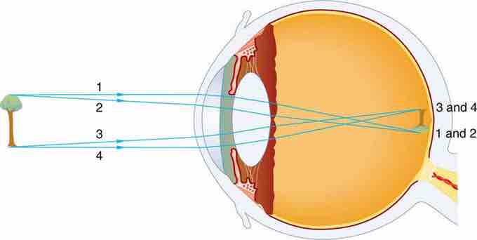
Vision Diagram
An image is formed on the retina with light rays converging most at the cornea and upon entering and exiting the lens. Rays from the top and bottom of the object are traced and produce an inverted real image on the retina. The distance to the object is drawn smaller than scale.
Eye Movement
Each eye has six muscles; lateral rectus, medial rectus, inferior rectus, superior rectus, inferior oblique, and superior oblique. All of these muscles provide differnt tensions and torques to control the movement of the eye. These are a few examples of types of eye movement:
- Rapid Eye Movement - Often referred to as REM, this happens in the sleep stage when most vivid dreams occur.
- Saccade - These are quick, simultaneous movements of both eyes, and is controlled by the frontal lobe of the brain.
- Vestibulo-ocular Reflex - This is the eye movement which is opposite to the movement of the head and keeps the object you are looking at in the center of vision.
- Pursuit Movement - This is the tracking movement when you are following a moving object. It is less accurate than the vestibulo-ocular reflex.