Intraembryonic Coelom Development
In the development of the human embryo the intraembryonic coelom (or somatic coelom) is a portion of the conceptus that forms in the mesoderm. During the second week of development the lateral mesoderm splits into a dorsal somatic mesoderm (somatopleure) and a ventral splanchnic mesoderm (splanchnopleure).
By the third week of development, this process gives rise to a cavity between the somatopleure and splanchnopleure referred to as the intraembryonic celom. This space later gives rise to both the thoracic and abdominal cavities.
Somite Development
In the developing vertebrate embryo, somites are masses of mesoderm that can be found distributed along the two sides of the neural tube. They will eventually become dermis (dermatome), skeletal muscle (myotome), vertebrae (sclerotome), and tendons and cartilage (syndetome).
The mesoderm found lateral to the neural tube is called the paraxial mesoderm. It is separate from the chordamesoderm underneath the neural tube. The paraxial mesoderm is initially called the unsegmented mesoderm in vertebrates, but is called the segmented mesoderm in chick embryos.
As the primitive streak regresses and the neural folds gather preceding the formation of the neural tube, the paraxial mesoderm divides into blocks called somites. Somites play a critical role in early development by participating in the specification of the migration paths of neural crest cells and spinal nerve axons.
Later in development, somites separate into four compartments:
Dermatome
The dermatome is the dorsal portion of the paraxial mesoderm somite. In the human embryo it arises in the third week of embryogenesis.
The dermatomes contribute to the skin, fat, and connective tissue of the neck and of the trunk, though most of the skin is derived from the lateral plate mesoderm.
Myotome
The myotome is that part of a somite that forms the muscles. Each myotome divides into an epaxial part (epimere), at the back, and a hypaxial part (hypomere) at the front.
The myoblasts from the hypaxial division form the muscles of the thoracic and anterior abdominal walls. The epaxial muscle mass loses its segmental character to form the extensor muscles of the neck and trunk of mammals.
Sclerotome
The sclerotome forms the vertebrae and the rib cartilage and part of the occipital bone. It forms the musculature of the back, the ribs, and the limbs.
Syndetome
The syndetome forms the tendons and some blood vessels.
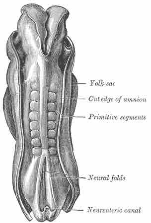
Somites
A dorsal view of a human embryo. The repetitive somites are marked with the older term primitive segments.