In the circulatory system, veins are blood vessels that carry blood towards the heart. Veins have thin, inelastic walls, and contain numerous valves in order to prevent backflow of blood. Most veins carry deoxygenated blood from the tissues back to the heart with the exceptions of the pulmonary and umbilical veins, both of which carry oxygenated blood to the heart.
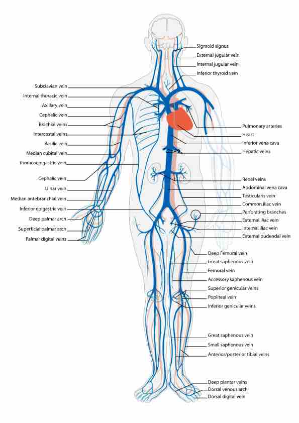
Principle Veins
This diagram shows the principal veins of the human body and their locations.
Veins can be broadly classified based on their depth within the body. Superficial veins are located close to the surface of the body and have no corresponding arteries, such as the great saphenous vein which runs the length of the leg. The deep veins lie deeper in the body and often run adjacent to corresponding arteries, such as the femoral vein which sits adjacent to the femoral artery in the thigh. Deep veins are often of larger caliber than superficial veins and carry the majority of the blood within the circulatory system. Communicating veins, or perforator veins if they pass through a large muscle mass, directly connect superficial and direct veins. The above veins form part of the systemic circulatory system. The pulmonary veins and venules that run from the lungs to the heart form part of the pulmonary circulatory system and are distinct from other veins in that they carry oxygenated blood.
Venae Cavae
The venae cavae are the veins with the largest diameter. Both enter the right atrium of the heart with the superior vena cava carrying blood from the arms, head, and thoracic cavity and the inferior vena cava carrying blood from the legs and abdomen. The inferior vena cava runs parallel to the abdominal aorta.
The superior vena cava is formed from the brachiocephalic veins which are in turn formed from the subclavian and internal jugular veins that serve the arm and head respectively. The inferior vena cava is formed from the common iliac veins that serve the legs and abdomen. The renal and hepatic veins from the kidneys and liver respectively also feed into the inferior vena cava.
Other Important Veins
Other important venous systems include the cardiac veins, which return blood from the heart tissue back to the general circulation. The cardiac veins merge into the coronary sinus, which empties directly into the right atrium.
The pulmonary veins are large blood vessels which receive oxygenated blood from the lungs and return it to the left atrium of the heart. There are four pulmonary veins, two from each lung, each of which forms from three to four bronchial veins. In approximately 25% of individuals, the left pulmonary veins may merge into a single vein; the same effect on the right side is only seen in approximately 3% of individuals.
The hepatic portal vein carries blood from the gastrointestinal tract to the liver. The portal vein is often described as a false vein because it conducts blood between capillary networks rather than between a capillary network and the heart. It functions to supply the liver with blood and required metabolites, but also ensures that ingested substances are first processed in the liver before reaching the wider systemic circulation.