The appendicular skeleton of vertebrates, including humans, consists of the bones that support and compose the appendages (for example, the arms and legs of humans). The word appendicular is the adjective of the noun appendage.
The appendicular skeleton includes the skeletal elements within the limbs, as well as supporting the pectoral and pelvic girdles.
The appendicular skeleton comprises 126 bones and is involved in locomotion and manipulation of objects in the environment. It is unfused, allowing for greater range of motion.
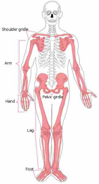
A diagram of the apendicular skeleton
Image depicting the human skeleton with the appendicular skeleton colored red.
Divisions of the Appendicular Skeleton
The appendicular skeleton is divided into six major regions:
- The pectoral girdles consist of 4 bones: The left and right clavicle (2) and the scapula (2).
- The upper arms and forearms are made up of 6 bones: The left and right humerus (upper arm, 2), the ulna (2), and the radius (forearm, 2).
- The hands have 54 bones: The left and right carpals (wrist, 16), metacarpals (10), proximal phalanges (10), intermediate phalanges (8), and the distal phalanges (10).
- The pelvis has 2 bones: The left and right hip bone (2).
- The thighs and legs have 8 bones: The left and right femur (thigh, 2), patella (knee, 2), tibia (2) and fibula (leg, 2).
- The feet and ankles have 52 bones: The left and right tarsals (ankle, 14), metatarsals (10), proximal phalanges (10), intermediate phalanges (8), and distal phalanges (10).
Pectoral Girdle
The bones of the pectoral girdle consist of two bones (scapula and clavicle) and anchor the upper limb to the thoracic cage of the axial skeleton.
The three regions of the upper limb are: arm (humerus), forearm (ulna medially and radius laterally), and the hand.
The base of the hand contains eight bones (carpal bones), and the palm is formed by five bones (metacarpal bones). The fingers and thumb contain a total of 14 bones, called phalanges.
Pelvic Girdle
The pelvic girdle is formed by a single bone, the hip or coxal bone, and serves as the attachment point for each lower limb. Each hip bone is joined to the axial skeleton by its attachment to the sacrum of the vertebral column. The right and left hip bones attach to each other anteriorly.
The lower limb contains 30 bones and is divided into three regions, the thigh, leg, and foot. These consist of the femur, patella, tibia, fibula, tarsal bones, metatarsal bones, and phalanges.
- The femur is the single bone of the thigh.
- The patella (kneecap) articulates with the distal femur.
- The tibia is located on the medial side of the leg,
- The fibula is the thin bone of the lateral leg.
The bones of the foot are divided into three groups, the tarsal bones, metatarsal bones, and phalanges of the foot.